- Diagnostic Imaging Vol 31 No 11
- Volume 31
- Issue 11
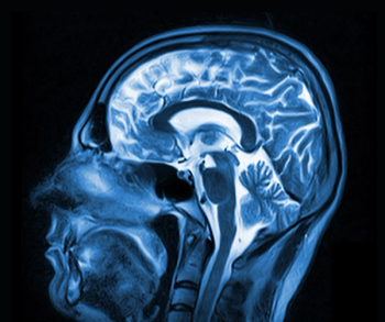
MEG finds source of ringing in ears
Researchers in Michigan are using magnetoencephalography of the brain to identify the source of tinnitus, a chronic ringing or buzzing in the ears or inside one's head.
Researchers in Michigan are using magnetoencephalography of the brain to identify the source of tinnitus, a chronic ringing or buzzing in the ears or inside one's head. They are also using MEG to map tinnitus’ site or sites for neurointervention and to assess the effectiveness of treatment.
According to the American Tinnitus Association, about 12 million people in the U.S. deal with tinnitus symptoms that require medical attention. Two million become disabled by it. The factors that trigger tinnitus have been identified, but its underlying causes remain unknown.
Some imaging modalities, like PET, SPECT, or fMRI, have been used to investigate its source in the brain, but none find where it originates, said principal investigator Dr. Michael D. Seidman, director of otologic and neurotologic surgery at Henry Ford Hospital in Detroit. MEG provides a much better diagnostic tool, he said.
“We now have a technique that gives us more pinpoint accuracy of where the sound of tinnitus may be being generated by the brain,” Seidman told Diagnostic Imaging.
Seidman and colleagues enrolled 17 patients with tinnitus and 10 normal individuals who underwent MEG for 10 minutes while sheltered from external auditory and visual stimuli. The researchers subjected the data to coherence analysis to measure synchronized neural activity.
They found that in patients with unilateral tinnitus MEG detected an area no larger than 2 mm in diameter in the auditory cortex of the brain hemisphere opposite to the perceived tinnitus. Patients with bilateral tinnitus showed activity in both brain hemispheres, but one side was dominant. Seidman released his group’s findings Oct. 3 at the 2009 American Academy of Otolaryngology–Head and Neck Surgery Foundation meeting.
Seidman has used MEG to map suspicious sites in the brains of six patients who underwent treatment with a low electrical current that “jams” the noise. He also used MEG to monitor symptom reduction after intervention. Four of those patients have experienced symptom relief. Results are positive enough to recommend routine clinical MEG use, Seidman said.
“We are excited about the technique,” he said. “Obviously more refinement needs to occur, but I would certainly encourage other centers that have MEG to use it.”
Articles in this issue
over 16 years ago
CMS accreditation could crimp in-office self-referralover 16 years ago
Nonphysician extenders can boost productivityover 16 years ago
Think outside the box and we'll clean up D.C.over 16 years ago
Radiologists in war-torn country reach out for global supportover 16 years ago
Just another day in the Baghdad Teaching Hospitalover 16 years ago
RIS/PACS serves as building block for electronic medical recordsover 16 years ago
Diagnostic Imaging at 30over 16 years ago
Canadian province probes new radiology reading messover 16 years ago
Winners get free mammograms!over 16 years ago
Ultrasound aids restaging of melanomaNewsletter
Stay at the forefront of radiology with the Diagnostic Imaging newsletter, delivering the latest news, clinical insights, and imaging advancements for today’s radiologists.


