Practical Insights on CT and MRI Neuroimaging and Reporting for Stroke Patients

In a recent video interview, Vivek Bansal, M.D., discussed a recent Radiology Partners publication that offers practical tips and best practice guidance on CT and MRI neuroimaging for stroke patients.
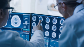
The recent Radiology Partners (RP) publication Neuroradiology and Stroke Clinical Pathway offers a practical best practice guidance for radiologists and technologists on the use and reporting of neuroimaging, including head and neck computed tomography angiography (CTA), cerebral perfusion CT and brain magnetic resonance imaging (MRI), for stroke patients.
In a recent interview, Vivek Bansal, M.D., makes it clear that the publication is geared toward achieving a consistent baseline standard for providing a high level of stroke imaging and care regardless of the size or resources of the given facility.
“We’re not rewriting the guidelines but (trying to) take some of these other hospitals or smaller practices, (facilities) with limited resources, or maybe don’t have neuroradiology specialty coverage, and bring everybody up to some baseline level to where we can say we’re giving optimal stroke care no matter where you go at any RP facility,” noted Dr. Bansal, a Houston-based neuroradiologist who serves as the national subspeciality lead for neuroradiology for Radiology Partners.
Emphasizing the constantly evolving nature and urgency of stroke care, Dr. Bansal said radiologists need to be active members of the stroke care team.
“In particular with stroke, we can’t be radiologists locked in our little reading rooms separate from everybody else and just putting out reports,” maintained Dr. Bansal. “We have to be part of the team … We have pushed to get really granular data and really help find the trouble points and fix them. That’s the key. When you do that, you get a lot of trust from your hospital system, and you can actually make processes better, and do better for the patients.”
(Editor’s note: To download the Neuroradiology and Stroke Clinical Pathway guidance,
For more insights from Dr. Bansal, watch the video below.
Newsletter
Stay at the forefront of radiology with the Diagnostic Imaging newsletter, delivering the latest news, clinical insights, and imaging advancements for today’s radiologists.


