
|Articles|August 26, 2008
Prostate MR tracks ultrasound ablation
High-intensity focused ultrasound ablation is used to manage localized prostate cancer after external-beam radiation therapy. But post-treatment alterations to prostate zonal anatomy hamper the assessment of local tumor progression that influences decisions about second-line treatment.
Advertisement
High-intensity focused ultrasound ablation is used to manage localized prostate cancer after external-beam radiation therapy. But post-treatment alterations to prostate zonal anatomy hamper the assessment of local tumor progression that influences decisions about second-line treatment. An interdisciplinary group from Sungkyunkwan University School of Medicine and Kangwon National University School of Medicine in South Korea tested two MR techniques for predicting local tumor progression.
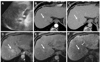
Twenty-seven patients underwent transurethral prostate resection and endorectal HIFU. These patients had clinical stage T1-T2 disease, no evidence of metastases, and a pre-ablation mean prostate-specific antigen level of 7.25 ng/mL. PSA levels continued to rise after treatment. Prostate MRI was done on a 3T system with two readers independently reviewing the MR images.
Based on the biopsy results, local tumor progression was found in 33% of the 162 prostate sectors. The diagnostic performance of dynamic contrast-enhanced MRI and diffusion-weighted imaging varied, with DCE-MRI delivering better sensitivity but DWI demonstrating better specificity. Accuracy rates for both readers and techniques were similar. False-positive results were higher for DCE-MRI than for DWI, the authors said. The areas under the receiver operator characteristic curves for reader 1 were 0.77 for both MRI techniques. The ROC curves for reader 2 were 0.85 for DCE-MRI and 0.81 for DWI.
In an interview with Diagnostic Imaging, study coauthor Dr. Chan Kyo Kim said DCE-MRI and DWI are part of the routine protocol for evaluating post-HIFU local tumor progression. Kim said that the urologists who performed the ablation procedures relied on imaging information as well as clinical and histopathological results.
Results were reported in the May issue of the American Journal of Roentgenology (2008;190[5]:1180-1186).
-By Shalmali Pal
Newsletter
Stay at the forefront of radiology with the Diagnostic Imaging newsletter, delivering the latest news, clinical insights, and imaging advancements for today’s radiologists.
Advertisement
Related Content
Advertisement

Latest CME


Advertisement
Advertisement
Trending on Diagnostic Imaging
1
Leading Breast Radiologists Discuss the Recent Lancet Study on AI and Interval Breast Cancer
2
Is AI Better Than Neuroradiologists at Evaluating Aneurysm Growth on CTA and MRA Scans?
3
FDA Clears AI-Powered Triage Platform for Digital Breast Tomosynthesis
4
FDA Clears 3T MRI Device for Neonates and Infants
5