|Videos|May 12, 2023
Emerging AI Advances in Cardiac Imaging
Author(s)Jeff Hall
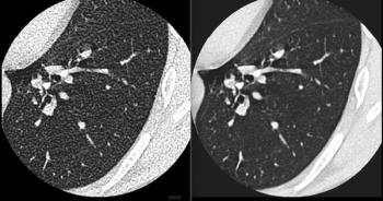
In a recent video interview, David Ouyang, M.D., shared insights from two recent studies he co-authored on the use of artificial intelligence (AI) to improve initial assessment of left ventricular ejection fraction (LVEF) on echocardiography and ascertain cardiac risks associated with changes in the left ventricle sphericity index seen on magnetic resonance imaging (MRI).
Advertisement
When evaluating an artificial intelligence (AI) model for initial assessment of left ventricular ejection fraction (LVEF) on echocardiograms, David Ouyang, M.D., said he was genuinely surprised that cardiologists had greater confidence in initial AI assessments than those provided by cardiac sonographers with an average of 14.1 years of experience.
In the randomized trial, recently published in
“We initially thought of the tool as kind of a streamlining tool where it speeds things up. We actually designed the trial as a non-inferior trial. We weren’t expecting AI to be better than sonographers. Hopefully, it would be equivalent,” noted Dr. Ouyang, who is affiliated with the Department of Cardiology at the Smidt Heart Institute and the Division of Artificial Intelligence in Medicine at the Cedars-Sinai Medical Center in Los Angeles. “We were pleasantly surprised that AI actually was superior. This really speaks to the high level of precision that automation delivers.”
(Editor’s note: For related content, see “
In a recent interview, Dr. Ouyang also discussed another
For more insights from Dr, Ouyang, watch the video below.
Advertisement
Related Content
Advertisement


Advertisement
Advertisement
Trending on Diagnostic Imaging
1
Should There Be Greater Oversight of MRI Safety?
2
Mammography Study: Can AI Detect Potential Breast Cancer Up to a Decade Prior to Diagnosis?
3
Thirteen Takeaways from New Report on AI in Health Care
4
The 2026 Medicare Shift in Radiology: What Changes, What to Do
5