
|Articles|May 10, 2012
Imaging Technique May Predict Sudden Cardiac Arrest Risk
Author(s)Marijke Vroomen Durning, RN
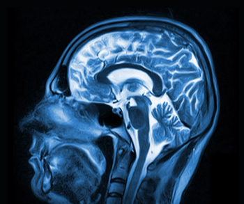
Imaging the loss of nerve function in the heart may better identify patients who would benefit from receiving implantable cardiac defibrillators (ICD) than current testing methods.
Advertisement
Imaging the loss of nerve function in the heart may better identify patients who would benefit from receiving implantable cardiac defibrillators (ICD) than current testing methods, said researchers from the University of Buffalo.
The researchers, who presented at the Heart Rhythm Society’s 33rd Annual Scientific Sessions, assessed patients by using PET imaging to quantify the patients’ amount of denervated myocardium, where sympathetic nerves in the heart have died or become damaged due to inadequate blood flow. Using cyclotron-generated radiopharmaceutical 11C-hydroxephedrine, they imaged heart’s ability to take up a radioactive tracer of norepinephrine, the neurotransmitter released from the heart's neurons.
Principal investigator, John M. Canty Jr., MD, a professor in the UB School of Medicine and Biomedical Sciences, and UB’s chief of cardiovascular medicine, explained that they wanted to develop an approach that could help identify who would be at higher risk of experiencing a sudden cardiac arrest.
“The principal question we posed with this study was whether the amount of denervated myocardium could predict sudden cardiac arrest,” he said. “We found that when at least 38 percent of the heart was denervated, there was a significant increase in the risk of cardiac arrest.”
These findings could change how it is decided who would receive an ICD, something that is currently done by measuring heart ejection fraction. Those who have an ejection fraction of 35 percent or less are considered candidates for ICDs.
Newsletter
Stay at the forefront of radiology with the Diagnostic Imaging newsletter, delivering the latest news, clinical insights, and imaging advancements for today’s radiologists.
Advertisement
Related Content


Latest CME


Advertisement
Advertisement
Trending on Diagnostic Imaging
1
Leading Breast Radiologists Discuss the Recent Lancet Study on AI and Interval Breast Cancer
2
Radiology Roundup of New FDA Clearances — February 1 — February 7
3
Is AI Better Than Neuroradiologists at Evaluating Aneurysm Growth on CTA and MRA Scans?
4
Diagnostic Imaging's Weekly Scan: February 1 — February 7
5