Diagnostic Imaging Europe
- Diagnostic Imaging Europe Vol 26 No 6
- Volume 26
- Issue 6
CR and DR triumph over conventional techniques
Digital radiography is now the standard technology for X-ray imaging. Computed radiography and solid-state (flat-panel) digital radiography are digital X-rays main detector systems.
Digital radiography is now widely regarded as the standard technology for x-ray imaging. Over the past 25 years, many screen-film systems have been replaced by digital units all over the world.
While modalities such as MRI and CT have been digital from the outset, projection radiography made the transition to digital relatively late. The eventual triumph of digital imaging over analog techniques was made feasible by improvements in technology. The switch to digital has also brought advantages in terms of image quality and improved image handling.
The physical properties of the various detector systems available offer competing advantages and disadvantages in terms of image quality, equipment handling, and cost.1,2 The image receptor material, readout process, and method of data processing can all differ from one detector system to another.
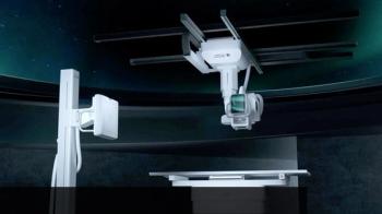
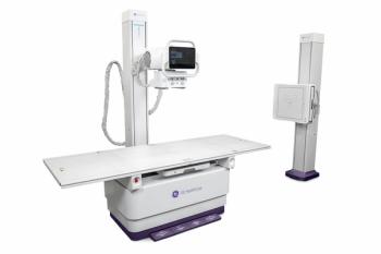
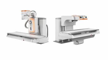
The two main detector systems used for digital x-ray are computed radiography (CR), based on storage phosphor plates, and solid-state (flat-panel) digital radiography (DR) (Figures 1 and 2).
INTRODUCING CR
CR was the first digital radiographic technique to be introduced. Systems are based on an imaging plate that is similar to the amplifier screen of a conventional radiography unit. The difference is that this storage phosphor imaging plate retains information from incident photons as a latent image. This image can then be retrieved following stimulation by a readout laser.
The first CR systems required more dose than screen-film systems to achieve a similar clinical performance. But these systems did allow the radiation dose to be varied. This meant that the dose could be reduced substantially in situations where reduced image quality was acceptable.3
Continuous improvements to the detector material and readout technology have led to substantial gains in dose efficiency and enhanced spatial resolution. Innovations include dual readout technology and needle-crystalline detectors. The dose efficiency of CR systems is now approaching that of DR systems, and is better than that of speed class 400 screen-film systems.1
DIRECT READOUT
Radiographic flat-panel DR detectors are characterized by a direct readout matrix of electronic elements. These elements are usually layers of amorphous silicon thin-film transistors (TFT), which are typically deposited on a piece of glass. This TFT layer is then coupled with an x-ray absorption medium.
There are two types of DR detector.3 Detectors that use a cesium iodide (CsI) or gadolinium oxysulfide (Gd2O2S) scintillator and light-sensitive photodiodes are called indirect-conversion TFT detectors or opto-direct systems.
The scintillators and phosphors used in indirect conversion detectors can be structured or unstructured. Unstructured scintillators scatter a large amount of light, reducing spatial resolution.4 Structured scintillators contain phosphor material in a needle-like structure, the needles being perpendicular to the screen surface. This increases the number of x-ray photon interactions and reduces the lateral scattering of light photons. The scintillator layer absorbs x-ray photons and converts them into fluorescent light. This light is then converted into an electric charge by means of an amorphous silicon photodiode array.5
Indirect conversion detectors are constructed by adding amorphous silicon photodiode circuitry and a scintillator to form the top layers in the TFT sandwich. These layers replace the x-ray semiconductor layer used in direct conversion devices (see below). The active area of the detector is divided into an integrated array of image elements (pixels). Each element contains a photodiode and a TFT switch available for the readout process.
Cesium iodide TFT systems are used widely for chest and skeletal radiography and are also suitable for real-time display. Mobile DR units that are appropriate for bedside use are also now available. These system use flexible substrates in place of glass, to make the detector more robust, and gadolinium oxysulfide as the scintillator.3
Direct conversion systems use amorphous selenium (a-Se) instead of amorphous silicon as the semiconductor material. This is due to the x-ray absorption properties and the extremely high intrinsic spatial resolution of amorphous selenium.6
An electric field is first applied across the selenium. X-ray exposure then generates electrons within the amorphous selenium layer. The absorbed x-ray photons are transformed into electric charges and drawn directly to the charge-collecting electrodes. Those charges generated (proportional to the incident x-ray beam) migrate vertically toward both surfaces of the selenium layer, without much lateral diffusion. Charges are drawn toward the TFT charge collector at the bottom of the amorphous selenium layer, where they are stored until readout. The charge collected at each storage capacitor is amplified and quantified to a digital code value for the corresponding pixel.
These systems are not suitable for real-time imaging due to their tendency to produce persistent latent images. They are used mostly in mammography units because they provide high dose efficiency for the frequency ranges needed in breast imaging.
EMERGING TRENDS
Mobility is an increasingly important topic in radiographic image acquisition. Portable DR detectors suitable for intensive care units and emergency departments, for instance, were introduced about five years ago. Early designs of portable detectors typically incorporated a gadolinium oxysulfide:terbium (Gd2O2S:Tb) intensifying screen and also utilized cable-transfer (i.e., not wireless) data.7
A new generation of portable detectors has been developed that provides wireless (WiFi) data transfer directly onto the departmental PACS network. These systems offer cable-free transfer of image data within seconds. For example, a WiFi-enabled portable detector is available as an add-on to Philips’ DigitalDiagnost DR system. This particular device has been designed to slot into a docking station that connects directly to the main system, which allows the detector battery to be recharged. The portable detector uses cesium iodide as the primary x-ray absorber and reportedly has a detector size of 35 × 43 cm with an image matrix size of 3000 × 2400 pixels. The pixel sampling interval is 144 µm (micrometers).
Another DR system of the future, Carestream’s DRX-1, is being tested now at our hospital (Figure 3). This digital radiographic image-capture system is used in conjunction with a conventional (analog) x-ray generation system from an original equipment manufacturer to produce digital projection radiographs of the human body. Data acquired by the detector are transmitted to the system console for storage, processing, display, and distribution to the healthcare facility network.
At the heart of the DRX-1 system is a thin flat-panel x-ray detector housed in a special cassette. It conforms dimensionally to the ISO 4090 standard and can be installed in an existing bucky as a direct and interchangeable replacement for existing CR or screen-film cassettes. The detector contains a full-field TFT readout array and integrated electronics for data storage and transmission. These can be powered by the DRX-1 detector’s internal rechargeable battery or through an electrical tether interface/cable connected to an external power supply.
Some groups are investigating alternative phosphor screen structures for use in indirect conversion detectors. One approach being considered is the integration of a pixilated superstructure of optically isolated cells into the cesium iodide layer to better suppress fluorescent light scatter.8 Another proposal is to pack powdered phosphors into a pixilated superstructure of cellular cavities. The walls of these microcavities would be made of a reflective material to prevent cross-talk between the pixels, minimizing blurring.9
Further improvements in DR detector performance may be achieved by replacing amorphous selenium with an alternative photoconductor. A number of polycrystalline materials are being evaluated for this role, including lead (II) iodide (PbI2), mercury(II) iodide (HgI2), lead (II) oxide (PbO), and cadmium-zinc-telluride (CdZnTe).10 These materials promise 50% to 100% greater x-ray absorption efficiency across the radiographic energy spectrum than an equivalent thickness of amorphous selenium, as well as a greater yield of signal electrons.
The above developments might conceivably lead to direct-conversion DR detectors that can compete with current indirect conversion detectors in terms of detective quantum efficiency (DQE) performance.7
In summary, CR and DR are mature technologies that have rapidly gained clinical acceptance. CR represents the older but highly versatile and economically attractive option that is equally suited to integrated systems and bedside cassette-based imaging.
DR detectors have essentially followed two different technological paths: indirect and direct conversion. Both offer superb image quality and options for dose reduction based on their high dose efficiency.11
Manufactures of solid-state DR detectors continue to refine their products and explore new technological themes. Imminent advances are expected in the realm of portable DR detectors. This innovation promises users greater operational freedom and improved workflow. Improvements to the physical characteristics of scintillator- and photoconductor-based x-ray absorbers are also being investigated, though certain technical difficulties must still be overcome.
Articles in this issue
almost 15 years ago
Intramedullary Spinal Sarcoidosis Myelopathyover 15 years ago
Marilyn Monroe's chest x-rays bring big moneyover 15 years ago
PET agent unlocks door to Alzheimer’s diseaseover 15 years ago
Endovascular embolization stops nosebleedsover 15 years ago
PET with CTC detects polyps well, requires no cleansingover 15 years ago
US identifies patients at higher stroke riskover 15 years ago
Algorithm lowers number of CTs for some strokesover 15 years ago
Letters to the Editorover 15 years ago
Vertebroplasty study adds weight, but not final wordover 15 years ago
Ultrasmall technology promises big boostNewsletter
Stay at the forefront of radiology with the Diagnostic Imaging newsletter, delivering the latest news, clinical insights, and imaging advancements for today’s radiologists.


