
|Articles|February 5, 2007
Contrast-enhanced ultrasound aids renal carcinoma evaluation
Author(s)Wendy Despain
Contrast-enhanced ultrasound offers a viable alternative to CT imaging for evaluating and monitoring renal cell carcinoma, according to three papers presented at the 2006 RSNA meeting.
Advertisement
Contrast-enhanced ultrasound offers a viable alternative to CT imaging for evaluating and monitoring renal cell carcinoma, according to three papers presented at the 2006 RSNA meeting.
Dr. Richard Barr, a professor of radiology at Northeastern Ohio University, and colleagues followed 234 patients beginning in 1998. Each patient had either an indeterminate renal mass or conflicting results from other imaging studies. The researchers used contrast-enhanced ultrasound considered state of the art at the time each patient presented, so several different doses and contrast agents, including Definity (Bristol-Myers Squibb) and Optison (Mallinkrodt), were used.
The researchers used their own vascularity algorithm to classify each mass as either benign or malignant based on blood flow patterns. Of the 234 cases, Barr and colleagues classified 179 as benign, 54 as malignant, and one as angiomyolipoma instead of renal cell carcinoma.
No lesions classified as benign by ultrasound have turned out to be malignant. Twenty were confirmed angiomyolipoma with other imaging techniques, 26 have not changed in three years, 15 have not changed in five years, and two resolved. Of the 54 lesions classified as malignant by ultrasound, 49 showed later pathological proof of malignancy. Results are not yet available on the rest, but contrast-enhanced ultrasound has so far performed with 100% sensitivity and specificity for classifying indeterminate renal cell carcinomas.
Another paper, presented by Dr. Michele Bertolotto from Trieste University in Italy, also assessed contrast-enhanced ultrasound in the diagnosis of atypical cystic renal masses.
Bertolotto and colleagues studied 51 patients with 54 atypical cystic renal masses identified and initially classified using CT. Twenty-two were classified as category II, 13 category IIF, 10 category III, and nine category IV. Ultrasound results using a modified Bosniak classification system agreed with CT classification on 87% of the lesions. Only seven lesions had different classifications based on ultrasound showing more septa than CT or upgrading wall thickness. The group recommends ultrasound as an alternative to CT for follow-up scans of complex cysts in patients with impared renal function.
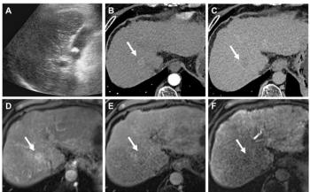
Finally, Dr. Franca Meloni of the radiology department at Ospedale Civile via Cesare in Milan and colleagues used contrast-enhanced ultrasound in follow-up care after radiofrequency ablation of renal cell carcinoma. They studied 24 patients with renal cell carcinomas ranging from 2.7 to 6 cm in diameter. Twenty-one had been judged inoperable, and three refused surgery. Twenty carcinomas were in the periphery, two were in the center of the kidney, and two had mixed locations. One month after the RFA procedure, and every six months after that, patients were assessed using multislice CT or MRI and contrast-enhanced ultrasound using a 2.4-mL intravenous bolus of a second-generation microbubble agent (SonoVue, Bracco).
Resulting images were evaluated at a consensus conference. CT showed six residual viable tumor foci out of the 24 cases. Five of these tumor foci were identified in the contrast-enhanced ultrasound images. Meloni and colleagues concluded that contrast-enhanced ultrasound is a viable technique for assessing the condition of renal cell carcinomas after RFA treatment, particularly for patients who will benefit from the absence of radiation exposure and lower toxicity of the contrast agent used in ultrasound.
Newsletter
Stay at the forefront of radiology with the Diagnostic Imaging newsletter, delivering the latest news, clinical insights, and imaging advancements for today’s radiologists.
Advertisement
Related Content
Advertisement


Latest CME


Advertisement
Advertisement
Trending on Diagnostic Imaging
1
Leading Breast Radiologists Discuss the Recent Lancet Study on AI and Interval Breast Cancer
2
Is AI Better Than Neuroradiologists at Evaluating Aneurysm Growth on CTA and MRA Scans?
3
Radiology Roundup of New FDA Clearances — February 1 — February 7
4
FDA Clears AI-Powered Triage Platform for Digital Breast Tomosynthesis
5