Diagnostic Imaging Europe
- Diagnostic Imaging Europe Vol 26 No 8
- Volume 26
- Issue 8
Breast tomosynthesis tackles new challenges
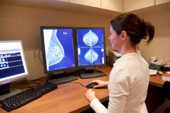
Mammography is the only screening modality that has been proven to reduce mortality from breast cancer.
Mammography is the only screening modality that has been proven to reduce mortality from breast cancer.1 The technique produces planar, projected images of the breast. Overlapping breast tissue on mammography’s projection views can, however, lead to lesions being obscured by overlaid parenchyma. Breast density may also affect the sensitivity of mammography.2
Unlike conventional mammography, which relies on the absorption of x-rays from a stationary tube to create a 2D projected image, digital tomosynthesis uses a moving x-ray source to generate 3D images. The technique is expected to overcome limitations related to superimposition of breast tissue, and thus is viewed as a promising adjunct to mammography.3,4
Although the basic principles of tomosynthesis have been known for many years, its development for mammography was hampered by the poor quality of available x-ray detectors. Advances in digital imaging, especially full-field digital mammography, have now allowed tomosynthesis to be implemented on clinical digital imaging units.
Systems for digital breast tomosynthesis are now available from several vendors, offering a range of angles and arc lengths. The motion of the tube may be linear, circular, or elliptical. The method of acquisition may be “step-and-shoot,” with one exposure taken at a series of discrete positions across the angular range, or continuous, where the exposure is pulsed throughout the motion of the x-ray source. Imaging may be performed with flat-panel detectors or multislit scanning systems. The multislit technique allows multiple projections to be acquired during a single scan, as the detector moves concurrently with the tube.5 The use of monochromatic x-ray sources has also been proposed.5
A set of low-dose source images is captured at various angles around the fulcrum during data acquisition while the breast is compressed. Images may be acquired in one or two views, typically without an antiscatter grid. Thin slices parallel to the detector plane are generated from the data sets to provide detailed visualization of the breast volume. Images are reconstructed using an algorithm usually similar to that used in CT reconstruction. Image data sets are sent from the acquisition workstation to the reading workstation.
A wide angular range allows thin-section reconstructions, which provides superior separation of sections. Depending on the range of the arc, between 30 and 80 sections, each 1 mm thick, can be obtained.
INITIAL EXPERIENCE
We performed breast tomosynthesis on a series of 150 patients who presented with clinical symptoms and whose mammograms or ultrasound scans revealed lesions categorized as BI-RADS 3, 4, or 5. All patients had craniocaudal (CC) and mediolateral oblique (MLO) digital mammography and tomosynthesis (MLO view) of the same breast. Ultrasound was performed at the discretion of the radiologist. Breast tomosynthesis was performed under protocols approved by the institutional review board. All women were at least 40 years old and all provided informed consent.
The tomosynthesis was performed on a prototype unit adapted from the Senographe DS (GE Medical Systems). Fifteen projection images were acquired over an angular range of 40º, using an acquisition time of 15 seconds. Acquisition parameters were selected manually for each patient according to a table indicating appropriate choices for a given breast density and breast thickness under compression. Resulting images were reconstructed in 1 mm increments using an iterative reconstruction algorithm.
Depending on technology, dose for one-view tomosynthesis was lower than the dose for two- to three-view digital mammography. Reconstructed images were viewed individually or in dynamic cine mode at a workstation. Maximum-intensity projections (MIPs) and average images were reconstructed from the thin slices.
MEETING EXPECTATIONS
Breast tomosynthesis is expected to improve cancer detection on several fronts.
Reduction of summation artifacts: Tomosynthesis can help differentiate superimposed tissue from real lesions, as expected. Figure 1 illustrates this, showing a hazy opacity on the MLO view that is not localized on the CC view. The opacity was still apparently visible on the localized view. Thin slices and reconstructed MIPs from tomosynthesis showed a normal parenchymal pattern of scattered fibroglandular tissue and no suspicious findings, obviating a biopsy in this case.
Mass characterization: The visibility of structures such as lesions in a selected cross section of breast tissue is better on tomosynthesis than on conventional mammography; the visibility of objects outside the selected section is reduced.6 Lesion boundaries consequently can be delineated more clearly on tomosynthesis images, a key point in the recognition of malignant lesions (Figure 2).
Multiple images: Additional lesions may be difficult to assess in dense breasts due to overlying breast tissue. Correlation between mammographic views and breast ultrasound may be challenging in the case of multiple masses. The 3D exploration offered by tomosynthesis may make it easier to depict and localize multiple images.
Lesion localization: Lesions may sometimes be seen only on one mammographic view (
Improved detection: Digital breast tomosynthesis provides better contrast between dense and less dense components, such as fibroglandular tissue and fat, a feature that may reveal lesions not visible on digital mammography. Contrast between lesions and surrounding parenchyma is also increased on tomosynthesis.
FFDM AND TOMOSYNTHESIS
Some authors have reported a better conspicuity of cancers and a more appropriate BI-RADS categorization when tomosynthesis is added to standard digital mammography. In one such study, 98 women presenting with abnormal screening mammography underwent breast tomosynthesis with one to three views.7 The image quality of tomosynthesis was equivalent (n = 51) or superior (n = 37) to diagnostic mammography in 89% of cases (88/99). For masses, tomosynthesis image quality was rated as equivalent in 26% (five/19) or superior in 68% (13/19) of cases to diagnostic mammography. Masses constituted 19% (19/99) of findings detected on screening mammograms, but were 35% (13/37) of findings in which tomosynthesis had superior image quality.
In another study, breast cancer visibility on digital tomosynthesis was compared with one- and two-view digital mammography in a series of 36 patients. Subjects were selected on the basis of their digital mammograms showing only subtle signs of cancer.8 Forty breast cancers were found. Visualization of 22 lesions was better with tomosynthesis when compared with single-view mammography. The BI-RADS classification was upgraded in 58% of those cases. Visualization of 11 lesions was clearer with tomosynthesis when compared with two-view mammography. Thirty-three percent of these lesions were reclassified upward.
FUTURE DIRECTIONS
Tomosynthesis is making great strides, but still faces challenges.
Detection: Dense breast parenchyma that obscures the borders of masses limits the detection of breast lesions, even with tomosynthesis. Single-view tomosynthesis may fail to detect lesions located deep within the breast, in common with mammography.
Calcifications: Their small size makes the depiction and morphological characterization of breast microcalcifications challenging for breast tomosynthesis. Calcification clusters were well detected in our experience. The morphological features of microcalcifications were, however, altered due to geometric parameters. Chen et al showed that arc-shaped projections can distort the shape of microcalcifications in breast tomosynthesis.9 Using some digital breast tomosynthesis algorithms, such as the traditional “shift-and-add” algorithm, the appearance of calcifications may be blurred in the direction orthogonal to the tube motion. The spatial distribution of microcalcifications within the breast could be assessed, particularly when using MIP reconstructions. Significant artifacts caused by large calcifications can be recognized easily.
Further evaluation of digital breast tomosynthesis is needed. Should it be performed routinely or as an adjunct to mammography? Should it be used for screening or only in a diagnostic setting? These questions remain unanswered. The optimal number of tomosynthesis views (one? two? three?) has yet to be defined. The optimal acquisition geometry, that is, the tomographic angle and number of projection views, as well as the optimal acquisition parameters, such as target filter, tube voltage, and exposure, are still being investigated. Multiple reconstruction algorithms and acquisition protocols have been compared.10,11 Initial reports on the use of tomosynthesis, as well as on CAD, are promising.12,13 Larger clinical trials are needed before these techniques can be widely used, however.
The combination of the 3D exploration of breast volume provided by tomosynthesis with contrast administration may afford in a single examination a detailed assessment of breast cancer morphological features and vascular enhancement kinetics. The interest in this technique lies in its potentially lower cost and wider availability compared with MRI. Preliminary experience with contrast-enhanced tomosynthesis has shown dual-energy and temporal subtraction techniques are feasible.14,15
The 3D visualization of the breast from digital tomosynthesis provides useful information relative to standard mammography. The clinical efficacy of this technique, however, requires further clarification. Additional investigation that will help define the situations best served by this new tool is currently under way.
Articles in this issue
about 15 years ago
After two decades, it's time to bid farewell and thanksabout 15 years ago
Popular Swiss chest specialist receives prestigious SFR awardabout 15 years ago
Sporting design wins Olympic coin contestabout 15 years ago
Critics line up to pour scorn on impact factorabout 15 years ago
Multislice CT urography characterizes renal TBabout 15 years ago
Breast MRI's role in mastectomies remains openabout 15 years ago
Dynamic volume propels CT into fourth dimensionNewsletter
Stay at the forefront of radiology with the Diagnostic Imaging newsletter, delivering the latest news, clinical insights, and imaging advancements for today’s radiologists.


