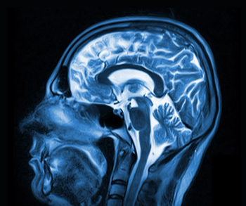
Brain MRI May Help Predict Outcome After Cardiac Arrest
Magnetic resonance imaging of the brain may help physicians predict patient outcomes following a cardiac arrest.
Determining MR imaging-based measures of the brain within two weeks of a patient’s cardiac arrest may help physicians predict patient clinical outcomes, according to a study published in
Researchers from the United States and France performed a prospective multicenter study to assess whether early brain functional connectivity is associated with functional recovery one year after cardiac arrest.
Forty-six patients who were comatose after a cardiac arrest were enrolled in the study. The principle outcome was cerebral performance category at 12 months. A favorable outcome was defined as cerebral performance category 1 or 2.
All participants underwent multiparametric structural and functional MR imaging less than 4 weeks (mean of 12.6 days) after their cardiac arrest. Using seed-based analysis of resting-state functional MR imaging data, within- and between-network connectivity was measured in:
• Dorsal attention network (DAN)
• Default-mode network (DMN)
• Salience network (SN)
• Executive control network (ECN)
Structural changes identified with fluid-attenuated inversion recovery and diffusion-weighted imaging sequences were analyzed by using validated morphologic scales. The association between connectivity measures, structural changes, and the principal outcome was explored with multivariable modeling.
The results showed at 12 months, 11 patients had a favorable outcome. Patients with favorable outcome had higher within-DMN connectivity and greater anticorrelation between SN and DMN and between SN and ECN compared with patients with unfavorable outcome, an effect that was maintained after multivariable adjustment. Anticorrelation of SN-DMN predicted outcomes with higher accuracy than fluid-attenuated inversion recovery or diffusion-weighted imaging scores.
The researchers concluded that using MR imaging to measure cerebral functional network connectivity obtained in the acute phase of CA were independently associated with favorable outcome at one year. The authors suggested that these findings warranted validation as early markers of long-term recovery potential in patients with anoxic-ischemic encephalopathy.
Newsletter
Stay at the forefront of radiology with the Diagnostic Imaging newsletter, delivering the latest news, clinical insights, and imaging advancements for today’s radiologists.


