
|Articles|November 6, 2008
Brain blood flow plays role in fibromyalgia symptoms
Author(s)Rebekah Moan
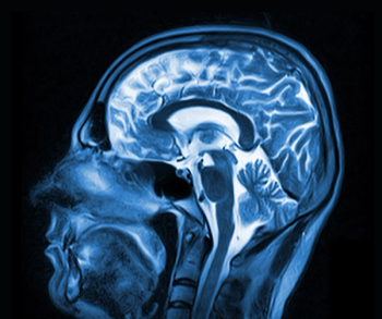
In a potential breakthrough study, French researchers used SPECT to identify brain abnormalities that present physiological evidence of fibromyalgia. The results quash the idea that diffuse, sometimes debilitating pain from the condition stems from anxiety and depression.
Advertisement
In a potential breakthrough study, French researchers used SPECT to identify brain abnormalities that present physiological evidence of fibromyalgia. The results quash the idea that diffuse, sometimes debilitating pain from the condition stems from anxiety and depression.
Fibromyalgia afflicts between three million and six million people in U.S., according to the National Institute of Arthritis and Musculoskeletal and Skin Diseases. Diagnosis is difficult because its symptoms overlap with many rheumatic conditions.
Dr. Eric Guedj of the Service Central de Biophysique et de Médecine Nucléaire at Assistance Publique des Hôpitaux de Marseille and colleagues investigated the specific clinical correlate of functional abnormalities of the brain using a technetium-99m-labeled probe and SPECT in 30 women (J Nucl Med 2008;49:1798-1803).
Compared with healthy control subjects, the 20 fibromyalgia patients exhibited posterior hyperperfusion in the somatosensory cortex and hypoperfusion of the frontal, cingulated, temporal, and cerebellar cortices. While also noting distinct perfusion in fibromyalgia patients, Guedj and colleagues found correlation between degree of perfusion and severity of pain.
Patients who experienced symptoms of fibromyalgia more intensely had higher blood flow to the parietal areas, which are known to discriminate pain intensity, as well as lower blood flow within anterior temporal areas, known to be involved with emotional aspects of pain.
Study subjects evaluated the severity of their disease symptoms using the Fibromyalgia Impact Questionnaire, a standardized, self-administered survey consisting of visual analog scales and questions regarding limitations on activities of daily living such as the number of days they were unable to work. Anxiety and depression were evaluated by the Hospital Anxiety and Depression Scale, a self-screening questionnaire.
The FIQ total score positively correlated with perfusion of the bilateral superior parietal lobule, bilateral precuneus, left postcentral cortex, and right pre- and postcentral cortex. The FIQ total score negatively correlated with perfusion of a left anterior temporal cluster, including the inferior, middle, and superior temporal gyri, fusiform gyrus, and uncus.
Patients were injected with cysteinate dimer labeled with technetium- 99m and kept at rest for one hour in quiet surroundings with their eyes closed. Guedj and colleagues performed SPECT using a dual-head gamma camera equipped with fanbeam collimation in 64 projections through 360° of rotation.
Previously, researchers thought functional brain abnormalities for fibromyalgia were related to psychological or psychiatric symptoms, Guedj said.
“We showed in our study that the functional abnormalities observed were mainly related to disability and not to anxiety and depression status,” he said.
The severity of fibromyalgia could be objectively assessed using brain imaging, according to Guedj.
“This parameter could be used to evaluate new treatment in clinical trials,” he said.
Limitations of the study include absence of a correlation between SPECT abnormalities and pain visual analog scale, the low number of participants, the difference in numbers between patients and the control group, and the usefulness of the questionnaires.
For more information from the Diagnostic Imaging archives:
Newsletter
Stay at the forefront of radiology with the Diagnostic Imaging newsletter, delivering the latest news, clinical insights, and imaging advancements for today’s radiologists.
Advertisement
Related Content
Advertisement


Latest CME


Advertisement
Advertisement
Trending on Diagnostic Imaging
1
Leading Breast Radiologists Discuss the Recent Lancet Study on AI and Interval Breast Cancer
2
Is AI Better Than Neuroradiologists at Evaluating Aneurysm Growth on CTA and MRA Scans?
3
FDA Clears 3T MRI Device for Neonates and Infants
4
FDA Clears AI-Powered Triage Platform for Digital Breast Tomosynthesis
5