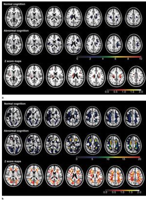
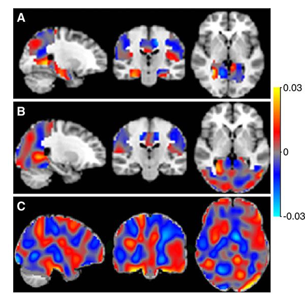


Functional MR imaging shows increased response in the brain after administration of low-dose methylene blue.
Functional MR imaging shows increased response in the brain after administration of low-dose methylene blue.

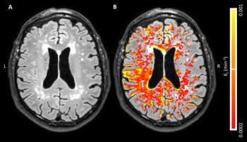
Using MRI to evaluate blood-brain barrier disruption following intracranial hemorrhage may predict severity after intervention.
Diffusion tensor imaging detected abnormalities in the brain among subjects with mild traumatic brain injury.
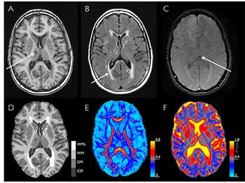
MRI may help detect Alzheimer’s before directly visible cerebrovascular abnormalities are present.

Use of brain imaging in the ED increased dramatically since 1994, according to a study at ACR 2016.
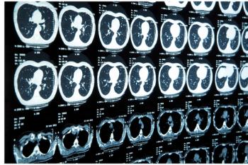
Brain CT and MRI for early stage lung cancer patients are not routinely recommended.

Radiologists detect changes in the brains of veterans who have sustained combat-related mild traumatic brain injuries.
Diffusion-tensor MRI algorithm may help physicians evaluate patients with mild traumatic brain injury and post-trauma migraines.

MRI distinguishes brain lesions, possibly supplementing existing diagnostic algorithms
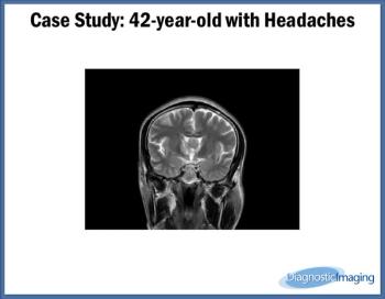
Case History: 42-year-old patient presents with complaints of headache.
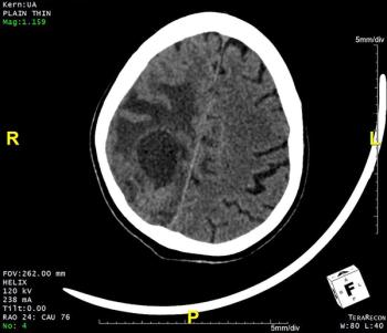
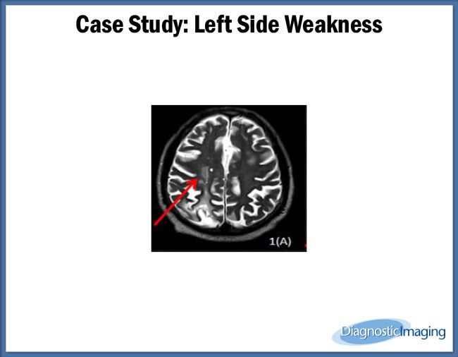
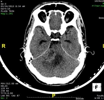
75-year-old male presented with left-sided weakness, headache, and altered sensorium.
MRI shows lasting brain damage among military personnel with MTBI from blast injuries.

Choosing Wisely forces neurology to confront its issue with overutilization in imaging.
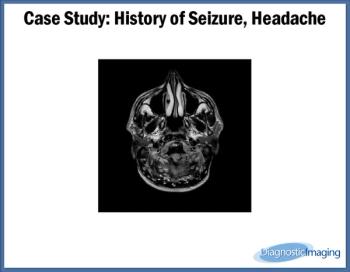
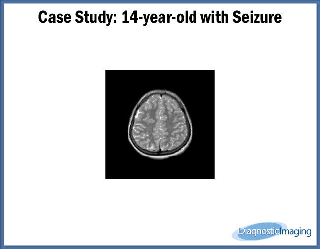
Case History: A 27-year-old patient presented with history of headache and seizure.
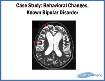
Case History: 60-year-old male presents with behavioral changes for 2 months in bipolar disorder.

MRI plus clinical criteria helps physicians determine which stroke patients would benefit from thrombectomy.
CHICAGO-MRI suggests that subclinical cardiac dysfunction could be indicative of early brain disease.

CHICAGO-MRI shows abnormal cerebral blood flow even if concussed athletes clinically ready to return to sport.
28-year-old male presented with sensorineural hearing loss for two months.
Using MRI to assess hippocampal grading, physicians may be able to predict Alzheimer’s disease up to seven years before dementia.

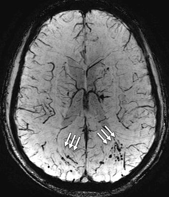
An earlier scan of a patient’s brain with traumatic brain injury is better for greater detection of microhemorrhages.

Use of MRI images may help psychiatrists determine effectiveness of antipsychotic drug treatment.
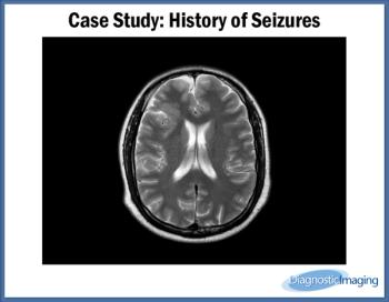
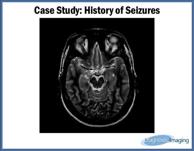
Case History: 35-year-old presents with history of seizures.
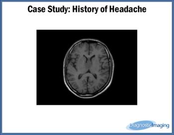
Case History: 40-year-old with history of headache.
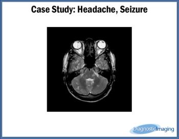
Case History: 24-year-old presents with complaints of headache and seizures.