


Case History: 49-year-old patient with large buccal mass, history of leakage through mass.
Case History: 49-year-old patient with large buccal mass, history of leakage through mass.
Computed tomography angiography helps physicians determine if patients with penetrating vascular trauma require surgery.

Case History: 65-year-old with lower left chest wall swelling since childhood.
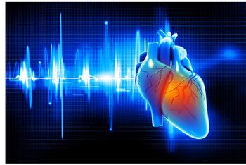
The combination of CT angiography and CT perfusion versus the combination of invasive coronary angiography plus single photo emission CT in predicting two-year MACE.

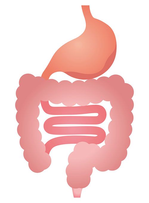
Rates of colorectal cancer screening via CT colonography are higher when patients have health insurance policies that cover the screening.

Case History: 62-year-old with history of breathlessness.

Using CT perfusion to track ovarian cancer treatment may help physicians determine prognosis.

Case History: 50-year-old patient with ovarian cancer and total abdominal hysterectomy presented with urinary incontinence.

Case History: 45-year-old patient presented with history of abdominal discomfort.
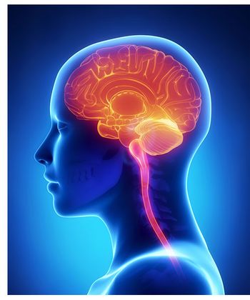
Using PET or CSF assay to detect amyloid plaques in asymptomatic adults may help detect future cognitive decline.
Case History: 50-year-old patient with ureteral stent evaluated with CT.
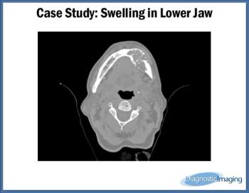
Case History: 60-year-old female presented with complaint of swelling of left mandible.
Radiographs can provide anatomic evaluation and appropriate starting point when investigating possible musculoskeletal infection.
Ventilation and perfusion scans to diagnose pulmonary embolism remain largely accurate.
Advanced imaging is being used more often in emergency rooms.
CT-Guided VAB outperforms prone stereotactic biopsy for fastest breast tissues biopsy.
Diagnostic reference levels help reduce radiation doses for CT examinations.
Combining FDG/PET with CT helps radiologists detect lymph node metastasis in high-risk endometrial cancer.
Using a comprehensive utilization management can help reduce the use of high cost imaging in primary care practices.
By adding a new category to lung CT reports, radiologists can identify more malignant lesions.
Radiation doses for CT scans may decrease when information is shared between institutions.
Image quality may be affected if reduced doses in 18F-FDG-PET/MRI are used for abdominal examinations.
Radiation doses for identical CT scans are still variable, despite lower levels overall.
Using weight-based protocol incorporating tube potential selection allows for lower volumes of iodinated contrast material in aortic CTA.
Using noninvasive techniques, such as CT angiography and CT perfusion, may help physicians identify patients at risk of major adverse cardiovascular events.