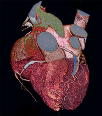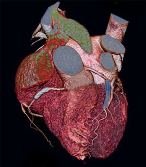
A 58-year-old man with a history of hypertension and hypercholesterolemia was admitted to the hospital with symptoms of suspected stable angina pectoris. The patient was referred to conventional coronary angiography after a positive exercise-ECG test. Conventional angiography showed significant stenoses at the level of the proximal right coronary artery (RCA) and the proximal left anterior descending coronary artery (LAD). Percutaneous intervention was undertaken and one bare-metal stent in the RCA and two overlapping bare-metal stents in the LAD were successfully implanted. The patient was referred to follow-up CT coronary angiography after 18 months.

