
|Articles|August 2, 2020
X-ray Signal Extraction Method Could Be Next Generation Breast Cancer Screening Technique
Author(s)Whitney J. Palmer
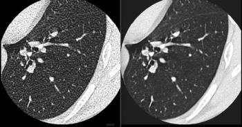
X-ray phase contrast imaging provides better soft tissue differentiation and tumor detection.
Advertisement
In a study published in
Although mammography is the conventionally preferred method for breast cancer screening, its limited image contrast mechanism can reduce the number of cancers it can identify. The technique called X-ray phase contrast imaging (XPCI) does offer better soft tissue differentiation and tumor detection, but the gold and silicon gratings used in it can reduce dose efficiency, meaning patients are exposed to more radiation.
To side-step this problem, a team led by Yongshuai Ge developed a CNN named XP-NET that was used to create a new XPCI signal extraction technique that could augment signal accuracy, leading to improved X-ray dose efficiency.
“We demonstrate that the deep convolutional neural network technique provides a promising approach to improve the grating-based XPCI performance and its dose efficiency in future biomedical applications,” Ge’s team wrote.
XP-NET’s special architecture design automatically performs XPCI signal retrieval and image quality enhancement in a sequence. By doing so, the CNN improved the phase signal accuracy by more than 15 percent compared to a conventional analytical method.
The team tested the CNN with both biological specimens and breast phantom studies, and they found that it was able to acquire phase images with half the dose, and the image quality was comparable to that acquired with the standard dose level.
These findings, the team said, point to the future potential low-dose pre-clinical uses of high quality breast X-ray phase contrast imaging.
Advertisement
Related Content
Advertisement


Advertisement
Advertisement
Trending on Diagnostic Imaging
1
FDA Clears AI-Powered Software for Improving Low-Contrast CT Detection
2
DeepHealth Launches AI-Powered Software for Radiology Reporting
3
Mammography Study: Can AI Detect Potential Breast Cancer Up to a Decade Prior to Diagnosis?
4
Thirteen Takeaways from New Report on AI in Health Care
5