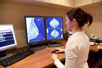
The State of CAD for Mammography
As computer-aided detection becomes the standard of care for mammography, how are radiologists using the tool, and what does its future hold?
Making screening mammography more effective has been on radiology’s to-do list for several years. One of the most commonly used tools to improve diagnostic sensitivity and specificity has been computer-aided detection. Over the past decade, the technology has made significant in-roads, but questions remain about how certain features should be used.
When paired with mammography, CAD helps radiologists identify any abnormal areas in breast tissue. To date, it’s been used mainly with 2D imaging, but work is underway to extend its utility to 3D imaging, as well.
Despite its maturation and widespread use, however, there isn’t yet consensus about whether CAD is truly useful. In fact, a 2011
The State of CAD Today
Even in the face of negative research, CAD has evolved over the past decade to be a nearly indispensable technology for the practicing radiologist, according to Bruce F. Schroeder, MD, medical director of Carolina Breast Imaging Specialists in Greenville, NC, and long-time breast imaging expert.
“If you’d asked me 10 years ago whether we needed CAD in mammography, I’d have said we weren’t sure what to do with it. It was new - we thought it had value, but that wasn’t proven,” Schroeder said. “Fast-forward to today, and it really is the standard-of-care. I don’t know of a practice that doesn’t use it.”
When used in conjunction with mammograms, CAD identifies and marks suspicious lesions and masses in breast tissue that radiologists could miss. The tool’s sensitivity substitutes for the human eye’s inability to focus on every pixel in an image. Consequently, he said, CAD catches the sub-millimeter calcifications radiologists can miss.
“We’re human, and we look at multiple images,” Schroeder said. “There is a fatigue factor involved, and we don’t always cover every location when looking at the image. [CAD] keeps you honest.”
CAD’s biggest benefit comes with correctly and objectively identifying and measuring breast density. This ability is significant, Schroeder said, because dense breast tissue can easily hide malignancies, leaving them undiscovered until they are untreatable.
“Human assessments of breast density are really subjective compared to the machine assessments that do volumetric measurements,” he said. “We’re really bad, as radiologists, at defining density. Show me the same film several times, and I’ll give you a different density.”
With 14 states - and more on the way - requiring radiologists to inform women whether they have dense breast tissue, CAD’s precision is invaluable. Not only does the accuracy minimize errors, but it also reduces needless fear among patients and unnecessary supplemental imaging.
CAD Challenges
But, as much as industry experts proclaim CAD to be the standard-of-care with mammography, evidence exists that providers aren’t relying on it to a large degree. With assistance from the Society for Breast Imaging and Diagnostic Imaging, Eliot Siegel, MD, diagnostic radiology professor and vice chair for research informatics at the University of Maryland, conducted a small survey of about 50 radiologists, asking how they actually used this technology. He and his colleague Baltimore-based Jonathan Mezrich, MD, JD, presented their findings at the 2013 Radiological Society of North America (RSNA) meeting.
As it turns out, Siegel said, providers are saying one thing and doing another.
“There’s really an interesting mismatch. Ninety percent of people told us they end up using [CAD], but only half of them say that they even sometimes rely on the findings. The other half: rarely or never,” Siegel said. “It’s an interesting dichotomy.”
Survey results also revealed 62 percent of respondents rarely or never changed their reading based on the CAD findings, and 36 percent said they rarely or never found the technology helpful. And, although 80 percent agreed CAD qualifies as the standard-of-care for screening mammography, 87 percent said radiologists would provide the same level of care they didn’t use CAD at all.
“This pattern leaves open to question the real level of importance CAD has,” Spiegel said. “It seems that mammographers are skeptical about its added value.”
Regardless of whether providers use CAD, there are real concerns about how they use it. If providers use CAD, are they obligated to follow its advice? If they don’t, are they vulnerable to a medical malpractice lawsuit? When saving images, should providers also save the CAD markings or can they discard them?
According to survey results, 72 percent of providers report they never or rarely save CAD markings, citing the belief that discarding them would decrease the chance of any future law suits. Not saving the CAD data, though, could be a mistake, Siegel said.
“There is a concept in law called spoliation that refers to the intentional withholding of evidence. In malpractice cases, if something is lost or destroyed that should’ve been in the medical record, the jury must presume the missing evidence is unfavorable,” he said. “In cases where CAD was used and the markings aren’t available, it could be considered spoliation.”
Siegel recommended radiologists always save CAD markings in case they ever need to prove the accuracy of their diagnoses. It can also be helpful as CAD technology advances, he said. In instances where law suits aren’t brought to court for years, an imaging record with markings from earlier-generation CAD technology can protect a provider if a cancer was missed.
He also cautioned radiologists to remember that CAD tools mark anything that appears abnormal, resulting in a high false-positive rate.
CAD’s Next Frontier
There’s wide consensus that CAD is now a mature technology with 2D imaging. Work is now underway to extend its utility to 3D imaging through tomosynthesis. Diagnostic, imaging, and surgical product vendor Hologic is leading the way with integrating these two tools. According to a company white paper, its ImageChecker CAD product is designed to increase identification of micro-calcifications in single tomosynthesis slices. Hologic currently has the only CAD tomosynthesis product on the market.
But the rest of the industry is also moving in this direction, said Stacey Stevens, senior vice president of marketing and strategy for advanced imaging analysis company iCAD. At November’s RSNA meeting, iCAD presented its developments with CAD and tomosynthesis as a “work in progress.”
CAD, Stevens said, will be even more valuable when used with 3D imaging than with 2D.
“When looking at 3D images, there’s much more data than with 2D. And, when you’re looking at 1-millimeter slices of the breast and there’s a tiny lesion, it’s very easy to miss it with that amount of information,” she said. “So, a big focus among vendors is to develop tomosynthesis CAD products that not only help increase sensitivity to finding cancer, but help significantly increase workflow.”
Although some radiologists report they use CAD even though they’re not fully convinced of its utility, Carolina Breast Imaging Specialists’ Schroeder said the industry is pivoting toward the next advance with this technology.
“The next frontier,” he said, “is going to be applying what we know in CAD to the 3D data sets that we’re now starting to see in clinical practice
Newsletter
Stay at the forefront of radiology with the Diagnostic Imaging newsletter, delivering the latest news, clinical insights, and imaging advancements for today’s radiologists.


