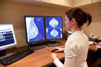
Report from NCBC: CAD boost in spotting cancers shows variation
There’s no doubt that computer-aided detection increases the ability to pick up breast cancers. But questions remain about which users benefit most from CAD, as cancer detection rates vary widely with breast imaging experience.
There's no doubt that computer-aided detection increases the ability to pick up breast cancers. But questions remain about which users benefit most from CAD, as cancer detection rates vary widely with breast imaging experience.
Even proponents note that improvements in the technology are needed to decrease false positives while increasing accuracy in detecting cancerous masses.
"Two excellent double readers are still better than the best CAD system," said Dr. Michael Linver, director of mammography at the Breast Imaging Center in Albuquerque. "But is this practical for most people in most centers? Probably not. CAD is the wave of the future."
Linver also cited CAD's caveats during a presentation Tuesday at the National Consortium of Breast Centers annual meeting in Las Vegas.
While published studies report a range of about 2% to nearly 20% in increased cancer detection, inexperienced breast imagers may benefit most from the technique. In a 2004 study involving 56,000 mammograms conducted at the University of Pittsburgh, overall cancer detection increased by only 1.7% (J Natl Cancer Inst 2004;96:185-190). That's much lower than previously published averages ranging from 7% at academic sites to 20% in the community setting.
A separate independent analysis of the Pittsburgh study found that among radiologists who read 8000 or more mammograms annually, cancer detection actually decreased with CAD. Low-volume readers, however, saw a nearly 20% increase in cancer detection, the same as that found in a community-based prospective study conducted in 2001. (Radiology 2001;220:781-786)
"The implication is that CAD has more value to those who don't read as many mammograms," Linver said. "In the U.S. today, more mammograms are being interpreted by low-volume readers, and this is an issue that CAD can address."
For CAD to increase its value to all users, several improvements are needed. CAD systems generate an average of two marks per study, Linver said. Taken in aggregate, a radiologist reviewing 1000 mammograms must investigate 2000 marks.
"That's a lot of marks, particularly when you consider that on average, there are five cancers for every 1000 patients," he said. "So for each cancer, I am looking at four hundred marks. That needs to be improved."
Higher cancer detection also means a concomitant rise in recall rates. And while CAD excels at picking up calcifications that radiologists may miss, systems are less expert at correctly identifying cancerous masses. CAD missed 33% of cancerous masses found by radiologists in the community-based study published in 2001.
CAD vendors are working to improve algorithms to address these problems, Linver said.
"CAD is increasing cancer detection. Hopefully, that means decreased morbidity and mortality, and even possibly decreased malpractice liability," he said. "But it does increase false positives, and reimbursement continues to be a problem with some payers."
Newsletter
Stay at the forefront of radiology with the Diagnostic Imaging newsletter, delivering the latest news, clinical insights, and imaging advancements for today’s radiologists.


