
|Articles|October 27, 2015
Rads Should Rely on Decision-Making, Not Detection, Skills
Author(s)Diagnostic Imaging Staff
Radiologists reading CT colonography may see difficult-to-detect polyps but not consciously register them.
Advertisement
Better decision-making improves reader performance of CT colonography, according to a study published in the
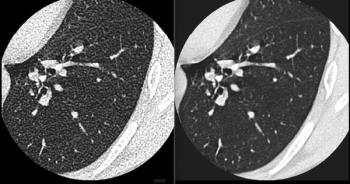
Researchers from the UK studied the eye-tracking of readers while they interpreted endoluminal CT colonography (CTC) studies. Their objective was to describe the characteristics of the polyps viewed but then dismissed incorrectly by radiologists, the eye movements during these errors, and the features provoking false-positive diagnoses.
Forty-two radiologists participated in the study. Seventeen were considered experienced (read more than 200 cases), nine were inexperienced (read fewer than 100 cases), and 16 were recruited from a UK training hospital. All were presented with 30 endoluminal CTC videos, each of which depicted a polyp. Half of the videos had computer-assisted detection (CAD), the other half did not. The researchers traced the radiologists’ eye movements while they were viewing the videos.[[{"type":"media","view_mode":"media_crop","fid":"42748","attributes":{"alt":"","class":"media-image media-image-right","id":"media_crop_6971303291111","media_crop_h":"0","media_crop_image_style":"-1","media_crop_instance":"4648","media_crop_rotate":"0","media_crop_scale_h":"0","media_crop_scale_w":"0","media_crop_w":"0","media_crop_x":"0","media_crop_y":"0","style":"height: 160px; width: 160px; border-width: 0px; border-style: solid; margin: 1px; float: right;","title":"©Alex Oakenman/Shutterstock.com","typeof":"foaf:Image"}}]]
Polyps were divided subsequently into “difficult to classify” and “easy to classify,” using a classification error threshold of more than 15%. Polyp diameter, height, and subjective conspicuity and the proportion of time viewed were compared between groups. The researchers successfully acquired eye-tracking data for 1,182 out of 1,260 case viewings (93.8%). The radiologists reported that difficult polyps, which had a mean diameter of 5.4 mm, were more difficult to notice, compared with the easier ones, which were a mean diameter of 8.2 mm.
The results showed that in 97% of the cases resulting in false-negative polyp diagnosis, the radiologist did initially observe the polyp. The polyps that were difficult to detect were subjectively less conspicuous (median score, 4) compared with the easier ones (median score, 2). “Readers spent proportionally less time viewing difficult polyps than viewing easy polyps (29.0 percent of the time they were on-screen versus 42.6 percent, respectively; p = 0.01) regardless of the presence of CAD,” the authors wrote.
The majority of polyps did attract reader gaze, meaning they were seen, but not necessarily noticed, the authors noted. The use of CAD did not change this. “Efforts to improve reader performance at CTC should focus on decision making rather than detection alone,” the concluded.
Advertisement
Related Content
Advertisement


Advertisement
Advertisement
Trending on Diagnostic Imaging
1
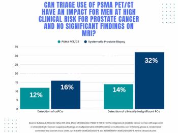
Study: PSMA PET/CT Reduces Biopsy Rate by Nearly 50 Percent for Men with Equivocal or Non-Suspicious Prostate mpMRI
2
Mammography Study: Can AI Detect Potential Breast Cancer Up to a Decade Prior to Diagnosis?
3
Breast Imaging in Focus: Can GLP-1 Receptor Agonists Reduce Breast Cancer Risk? What New Research Reveals
4
The Hidden Social Price of Remote Work in Radiology
5