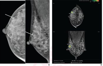
Radiologists may be able to predict the presence of ductal carcinoma in situ (DCIS) and invasive cancer with fairly high accuracy, according to a study published in the American Journal of Roentgenology. Researchers from California, Florida, and Texas sought to determine radiologists' estimated percentage likelihood assessments for the presence of DCIS and invasive cancer may predict histologic outcomes. The researchers retrospectively reviewed 250 cases that had been categorized as BI-RADS category 4 or 5 at four University of California Medical Centers. Ten academic radiologists who had been practicing from one to 39 years participated. Readers assigned BI-RADS category (1, 2, 3, 4a, 4b, 4c, or 5), estimated percentage likelihood of DCIS or invasive cancer (0 to 100 percent), and confidence rating (1 = low, 5 = high) after reviewing screening and diagnostic mammograms and ultrasound images. ROC curves were generated. The results showed that 62 percent (156/250) of lesions were benign and 38 percent (94/250) were malignant: Number of lesions AUC valuesDCIS 26 (10%) 0.731–0.837Invasive cancers 20 (8%) 0.830–0.907DCIS plus invasive cancers 48 (19%) - The sensitivity of 82 percent (56/68), specificity of 84 percent (153/182), positive predictive value (PPV) of 66 percent (56/85), negative predictive value (NPV) of 93 percent (153/165), and accuracy of 84 percent ([56 + 153]/250) were calculated using an estimated percentage likelihood of 20 percent or higher as the prediction threshold for invasive cancer for the radiologist with the highest AUC. Every 20 percent increase in the estimated percentage likelihood of invasive cancer increased the odds of invasive cancer by approximately two times, the authors wrote. For DCIS, using a threshold of 40 percent or higher, sensitivity of 81 percent (21/26), specificity of 79 percent (178/224), PPV of 31 percent (21/67), NPV of 97 percent (178/183), and accuracy of 80 percent ([21 + 178]/250) were calculated. Similarly, these values were calculated at thresholds of 2 percent or higher (BI-RADS category 4) and 95 percent or higher (BI-RADS category 5) to predict the presence of malignancy. The researchers concluded using likelihood estimates, radiologists may predict the presence of invasive cancer with fairly high accuracy. Radiologist-assigned estimated percentage likelihood can predict the presence of DCIS, albeit with lower accuracy than that for invasive cancer.


