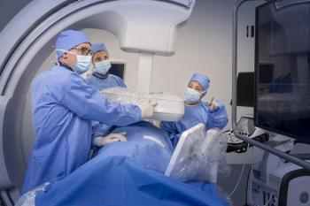
Optical technology sheds light on prostate imaging
Imulux, a pioneer in optical coherence tomography, showcased its FDA-cleared technology, Niris, at the World Congress of Endourology in Cleveland last week. Unlike other optical imaging tools that focus on the breast or brain, Niris renders images of the prostate. The system achieves a spatial resolution of 0.01 mm, which is well beyond the reach of diagnostic ultrasound.
Imulux, a pioneer in optical coherence tomography, showcased its FDA-cleared technology, Niris, at the World Congress of Endourology in Cleveland last week. Unlike other optical imaging tools that focus on the breast or brain, Niris renders images of the prostate. The system achieves a spatial resolution of 0.01 mm, which is well beyond the reach of diagnostic ultrasound.
About 3000 patients have undergone exams with Niris at 27 clinical sites around the world. Imulux has sold 12 products and loaned out another 15, but the company is only beginning its marketing effort. First Imulux strategists want to gather data to support the clinical value of the product, which cleared the FDA nearly two years ago.
"We are talking about changing the way medicine is practiced in terms of how we hope the product will be applied," said Jerry Cirino, CEO of Cleveland-based Imulux. "Physicians are bombarded with new things all the time. Getting a share of their minds is something we will be focusing on."
Imulux is encouraging users to publicize their clinical findings in presentations and publications. If interest rises, it will be met by Imulux in the U.S. and its distributors signed to handle China, Australia, and Korea. Another distributor is close to signing in Japan.
Priced at about $65,000, Niris might be amortized over relatively few procedures performed in a single year. The challenge is obtaining reimbursement.
Opthalmological applications are reimbursed, but dedicated CPT codes do not exist. Reimbursement is determined on a case-by-case basis. In the U.S., the company advises that prior authorization be obtained and submitted to the insurer to guarantee payment.
The potential exists for Niris to accomplish great things, according to Cirino. "It can visualize disease in the earliest stages of pathology," he said. "It is performed without contrast, at the point of care, and without the need for a specialist's review."
Niris is built on research conducted by scientists at the Russian Academy of Sciences, specifically at its Institute of Applied Physics. The researchers behind its development traded their rights to the intellectual property for shares in privately held Imulux. The current form of the technology provides 2D, cross-sectional images of prostate tissue, but its applications depend primarily on the form of delivery.
Optical coherence technologies such as Niris might be used to image any epithelial surface, including the skin, GI tract, colon, and urinary tract. It can be driven to other structures in the body using a laparoscope, according to Dr. Michael J. Manyak, a urologist at the Center for Prostate Disease Research at Walter Reed Army Medical Center, who has ties to Imulux.
In preliminary research, Manyak found that Niris could be used to identify the neurovascular bundles that must be preserved when performing prostatectomy to maintain erectile function. These bundles are difficult to see with conventional technologies, he said, but they are clearly apparent with the Niris system.
Similarly, Niris has proved potentially useful in determining the depth of penetration of bladder cancer. This is a critical issue in determining a patient management strategy, Manyak said.
"Once cancer penetrates into the muscle, you have a very aggressive cancer and you have to remove the entire bladder," he said.
On an even more fundamental level, Niris images may provide the basis for determining whether suspicious tissue is cancerous, thereby reducing, if not eliminating, the need for biopsy.
The name Niris reflects the source of its diagnostic power, near-infrared (NIR) light. It uses a lateral scanning mechanism embedded in a thin probe to visualize tissues about 2 mm from the transmitter. The scatter of light reveals tissue microstructure that may be valuable in assessing abnormal tissue.
The device consists of an imaging console about the size of a desktop computer and a detachable, flexible fiberoptic probe with a 2.7-mm outer diameter and length of two to five meters. The probe's narrow diameter allows it to be inserted into the working channel of rigid endoscopes, according to Cirino. Engineers are now trying to reduce the size further to allow it to fit into smaller, flexible cystoscopes.
"This will be part of our marketing strategy as we take the Niris system into the urology sector," he said.
During operation, near-infrared light is transmitted from the Niris console through the probe's optical fiber into the patient's tissue. The backscatter, collected by the probe, produces a high spatial resolution image, scanning laterally across the tissue surface. Scan time is about one second. The computer combines data obtained from in-depth and lateral scanning to create a 2D, cross-sectional image.
The company acknowledges that users must complete a learning curve to be able to interpret the images, but specialized training in radiology is not necessary. The system is designed for use by a physician or other medical personnel at the point of care. Efficient review and interpretation of Niris images depends on the user's experience with the system, according to the company.
"We have had physicians who were very comfortable using this system after 30 minutes of training," Cirino said.
Newsletter
Stay at the forefront of radiology with the Diagnostic Imaging newsletter, delivering the latest news, clinical insights, and imaging advancements for today’s radiologists.


