
|Articles|September 22, 2008
MRI reveals inner ear anomalies in children with hearing loss
Author(s)NeedsFixing
A study involving the imaging histories of more than 200 children with sensorineural hearing loss recommends MRI instead of CT for identifying soft-tissue defects associated with inner ear anomalies.
Advertisement
A study involving the imaging histories of more than 200 children with sensorineural hearing loss recommends MRI instead of CT for identifying soft-tissue defects associated with inner ear anomalies.Sensorineural hearing loss affects thousands of children per year, according to principal investigator Dr. John E. McClay. About half of all cases are thought to be genetic, 25% are acquired, and 25% originate from unknown causes.Radiography, including plain-film x-rays and CT, is often used to evaluate inner ear abnormalities in children with hearing loss. These methods evaluate the bones that contain the working components of inner-ear hearing. However, defects in the soft tissue within these bone structures also may be responsible for hearing loss.McClay and colleagues at the University of Texas at Southwestern Medical Center and Children's Medical Center Dallas analyzed the medical records of 227 children aged one month to 17 years (average age 5.3 years) with a diagnosis of sensorineural hearing loss. The children underwent MRI between June 1996 and June 2002. A total of 170 children had clinical information available and were included in the study. Of these, 101 (59%) had hearing loss in both ears and 69 (41%) had hearing loss in one ear, adding up to a total of 271 ears with sensorineural hearing loss.The results were published in the September issue of Archives of Otolaryngology -- Head & Neck Surgery (2008;134[9]:945-952).
The MRI results showed the following:
- 108 ears (40%) had inner ear abnormalities.
- 87 (32%) had abnormalities of the cochlea, a spiral structure containing hair cells integral to hearing: 63 (23%) with mild abnormalities and 24 (9%) with abnormalities considered moderate to severe.
- 49 ears (18%) had either missing (26, or 53%) or deficient (23, or 47%) cochlear nerves.
- Ears with severe and profound hearing loss had more abnormalities than those with mild and moderate hearing loss (48% versus 29%).
- Children with moderate, severe, or profound hearing loss in one ear had more inner ear abnormalities than children with hearing loss of the same severity in two ears (62% versus 38%).
McClay and colleagues concluded that a thorough workup to identify the cause of sensorineural hearing loss should be considered in each patient, though the specific origin of sensorineural hearing loss may remain undiagnosed in many patients.
High-resolution CT has been the imaging modality of choice in the initial workup of these patients, McClay said. The study indicates, however, that soft-tissue structures of the inner ear responsible for the electrochemical transfer of sound to the brain, such as the membranous labyrinth and the cochlear nerve, are not evaluated well with that modality. "With MRI, these soft-tissue components of hearing from the cochlea to the auditory cortex can be elucidated, which should improve our ability to appropriately diagnose the location of the defect in these children with sensorineural hearing loss," he said.
Advertisement
Related Content
Advertisement


Advertisement
Advertisement
Trending on Diagnostic Imaging
1
The Hidden Social Price of Remote Work in Radiology
2
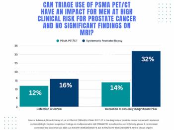
Study: PSMA PET/CT Reduces Biopsy Rate by Nearly 50 Percent for Men with Equivocal or Non-Suspicious Prostate mpMRI
3
Diagnostic Imaging's Weekly Scan: June 7 — June 13
4
Addressing Challenges in Radiology Reporting
5