
|Articles|September 15, 2020
MRI Knee Protocol Halves Scan Time, Safeguards Imaging Quality
Author(s)Whitney J. Palmer
The Compressed SENSE MRI technique can reduce scan time by more than half without impacting image quality or diagnostic certainty.
Advertisement
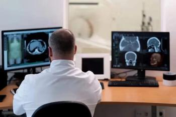
Radiology practices can cut the time it takes to conduct knee MRI scans by roughly half with a new protocol that does not create any negative impacts on image quality or diagnostic certainty.
By using Compressed SENSE (CS) to truncate 2D and 3D fat-saturated sequences of the knee, investigators from the University Hospital of Cologne in Germany succeeded in reducing exam times. Doing so can potentially help practices control their costs and improve their throughput. they said.
The team, led by Andra-Iza Iuga, a radiology resident doctor at the University Hospital, published their findings in the Sept. 10
Related Content:
These results clearly show, the team said, that using CS acceleration offers faster exams while leaving image quality and diagnostic certainty untouched. It also offers other benefits for the patient.
“Optimization of scan times not only increases patient’s comfort and decreases the vulnerability of motion artifact,” the team explained, “but it also represents a key factor for cost reduction of MRI exams as it reduces the fixed costs per examination.”
As a technique, CS takes less time because it gathers less sample data for each pixel. To determine how well the technique actually works, Iuga’s team recruited 20 healthy volunteers to test it with a 3T scanner. Each patient underwent a standard protocol MRI, as well as additional scans that used a variety of accelerating factors. Three independent readers examined and rated the images for signal-to-noise, contrast-to-noise, root-mean-square error, and structural similarity index.
According to their analysis, the team determined scan times decreased with an increasing CS factor. Based on their results, the 2D standard sequences was rated the best for diagnostic certainty and overall image impression.
While these findings point to CS efficacy, the team said, additional work is needed.
“Future investigations should consider larger samples sizes and use patients to analyze different knee pathologies and transferability to clinical routine,” they said.
Advertisement
Related Content


Advertisement
Advertisement
Trending on Diagnostic Imaging
1
FDA Approves Low-Dose MRI Contrast Agent Gadoquatrane
2
Study: PSMA PET/CT Reduces Biopsy Rate by Nearly 50 Percent for Men with Equivocal or Non-Suspicious Prostate mpMRI
3
The Hidden Social Price of Remote Work in Radiology
4
Molecular Imaging in Focus: Heather Jacene, MD, Charts Priorities as the New President of SNMMI
5