MRI Detects Brain Alterations in Epilepsy Beyond Temporal Lobe
Diffusion tensor imaging has detected that alterations in brain connections among patients with temporal lobe epilepsy are more widespread than thought.
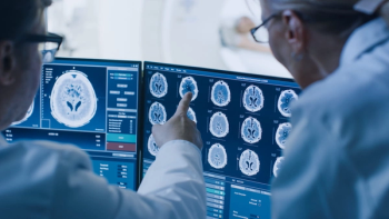
Diffusion tensor imaging detects alterations in brain connections in patients with temporal lobe epilepsy (TLE) beyond the temporal lobe, according to a study published online in the journal Radiology.
Researchers from Massachusetts General Hospital and Brigham and Women’s Hospital in Boston sought to evaluate if diffusion tensor imaging would help identify brain connection alterations among 24 patients, (age range from 12 to 65 years, mean age 33), with left TLE and 24 healthy controls (age range 19 to 55 years, mean age 25). Sixty-direction diffusion-tensor imaging and magnetization prepared rapid acquisition gradient-echo (MP-RAGE) magnetic resonance imaging volumes were analyzed.
The researchers found that structural connectivity was decreased throughout the default mode network (DMN) among the patients with TLE, compared with the healthy controls. The patients exhibited a decrease in long-range connectivity of 22 percent to 45 percent. “Using diffusion MRI, we found alterations in the structural connectivity beyond the medial temporal lobe, especially in the default mode network,” co-author Steven M. Stufflebeam, MD, said in a release.
The patients also had an 85 percent to 270 percent increase in local connectivity within and beyond the DMN. The researchers believe that this may be an adaptation to the loss of long-range connections.
More research is needed, the authors acknowledged. “Our long-term goal is to see if we can predict from diffusion studies who will respond to surgery and who will not,” said Stufflebeam.
Newsletter
Stay at the forefront of radiology with the Diagnostic Imaging newsletter, delivering the latest news, clinical insights, and imaging advancements for today’s radiologists.


