
|Articles|July 1, 2005
MR spectroscopy moves from the brain to bones
Characterizing musculoskeletal tumors with MR spectroscopy could eliminate the need for standard imaging and histological staging. Researchers at Johns Hopkins University have found preliminary success using MRS at 3T to diagnose bone tumors.
Advertisement
Characterizing musculoskeletal tumors with MR spectroscopy could eliminate the need for standard imaging and histological staging. Researchers at Johns Hopkins University have found preliminary success using MRS at 3T to diagnose bone tumors.
Dr. Laura M. Fayad, an assistant professor of radiology, and colleagues examined six bone tumors. They correctly detected a choline peak in two malignant cases and no choline peak in four benign cases, Fayad told Diagnostic Imaging.
The group had previously been successful in using MRS at 1.5T to investigate resected tumors. Fayad reported that study at the 2004 RSNA meeting.
Advertisement
Related Content
Advertisement


Advertisement
Advertisement
Trending on Diagnostic Imaging
1
What New Phase 3 MRI Findings Reveal About Survodutide in Patients with Obesity and MASLD
2
PSMA PET Plays Key Role in Multinational Phase 3 Trial of Perioperative Apalutamide for PCa
3
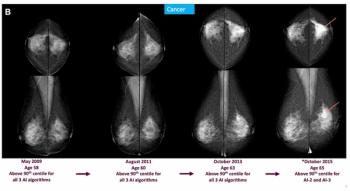
Mammography Study: Can AI Detect Potential Breast Cancer Up to a Decade Prior to Diagnosis?
4
The 2026 Medicare shift in radiology: What changes, what to do
5