
|Articles|June 25, 2020
Imaging Reveals Emerging Post-COVID Disease in Children
Author(s)Whitney J. Palmer
Multisystem Inflammatory Syndrome in Children appearing in pediatric patients with COVID-19 infection and exposure.
Advertisement
Imaging findings from several modalities have revealed an emerging disease in children who either have COVID-19 infection or who have been exposed to the virus, according to a new case series.
Known as Multisystem Inflammatory Syndrome in Children (MIS-C), this condition is characterized by airway inflammation, rapid development of pulmonary edema, coronary artery aneurysms, and extensive intra-abdominal inflammatory changes. A team of investigators from Evelina London Children’s Hospital in London assessed these findings in children admitted to the hospital and published their findings in the June 25
“Our hospital saw an unprecedented cluster of children presenting with MIS-C, a new hyperinflammatory syndrome in children related to the current COVID-19 pandemic – the recognition of which led to a national alert,” said lead study author Shema Hameed, M.B.B.S., consultant pediatric radiologist at Evelina London Children’s Hospital. “Our intention is to bring these findings to the attention of the wider radiological community.”
Initially, she said, the children’s symptoms, including fever, headaches, abdominal pain, rash, and conjunctivitis, potentially pointed to a diagnosis of Kawasaki disease, a condition that causes blood vessel wall inflammation, Kawasaki-disease shock syndrome, or toxic-shock syndrome. However, the symptoms were atypical and more severe.
To determine what was actually going on, the researchers performed a retrospective review of clinical, laboratory, and imaging findings, including ultrasound, CT, and X-ray studies, on the first 35 children under age 17 who were admitted to the pediatric hospital and who met the MIS-C case definition. The group, admitted between April 14, 2020, and May 9, 2020, included 27 boys and eight girls with an average age of 11.
Symptoms
Upon analysis of the medical data, the investigators identified several common symptoms. The most frequently occurring was fever, present in 94 percent of children. In addition, gastrointestinal symptoms, including abdominal pain, vomiting and diarrhea were seen in 86 percent of children, rash was present in 37 percent, and conjunctivitis developed in 26 percent. Of the group, 60 percent were in shock.
In many cases, children required substantial medical intervention. According to the team, 69 percent of patients had a clinical status severe enough to warrant management in the pediatric intensive care unit. Twenty percent of those children needed mechanical ventilation, and 57 percent required inotropic support. Due to severe myocardial dysfunction, two children needed extracorporeal membrane oxygenation, and lab tests revealed that all children had abnormal white blood cell counts.
Imaging Findings
Overall, the investigators identified a pattern of imaging findings that indicated post-COVID-19 MIS-C. Based on chest X-ray, chest CT, and abdominal ultrasound, they pinpointed airway inflammation, rapidly progressive pulmonary edema, coronary artery aneurysm, and extensive abdominal inflammatory changes within the right iliac fossa.
For modality-specific results, the investigators found:
Chest X-ray: All 35 children underwent this study based on fever, sepsis, or multi-system inflammation features. Of the group, 19 X-rays were abnormal with the most common finding being bronchial wall thickening.
Chest CT: Conducted in three children, the most common findings were basal consolidation and collapsed lung with pleural effusions.
Abdominal ultrasound: Five children underwent these studies, and findings included inflammatory changes within the right iliac fossa with mesenteric fat stranding, lymphadenopathy, and bowel wall thickening, along with free fluid identified in the pelvis.
Cardiac CT: Echocardiogram findings revealed cardiac dysfunction, including deteriorating myocardial function, myocarditis, pancarditis, pericardial effusions, and coronary artery aneurysms, in 18 children. Thirty patients underwent cardiac CT with six showing coronary artery aneurysms.
Investigators acknowledged that their study population was small, limiting the ability to make their findings generalizable. Consequently, they outlined the need for further investigations. But, these results are still valuable in adding knowledge to the understanding of how COVID-19 affects children.
“With these detailed findings and our postulated mechanisms for them, we hope to raise the awareness of this emerging condition among a radiology readership,” they said.
Advertisement
Related Content
Advertisement


Advertisement
Advertisement
Trending on Diagnostic Imaging
1
FDA Clears AI-Powered Software for Improving Low-Contrast CT Detection
2
DeepHealth Launches AI-Powered Software for Radiology Reporting
3
Mammography Study: Can AI Detect Potential Breast Cancer Up to a Decade Prior to Diagnosis?
4
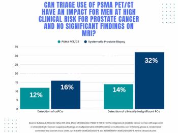
Study: PSMA PET/CT Reduces Biopsy Rate by Nearly 50 Percent for Men with Equivocal or Non-Suspicious Prostate mpMRI
5