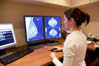
Imaging provides follow-up after breast cancer treatment
Many women with early-stage breast cancer undergo successful treatment for the disease. Some women who have been treated, however, may develop recurrent cancers. Imaging surveillance can detect the recurrence in time for treatment.
Many women with early-stage breast cancer undergo successful treatment for the disease. Some women who have been treated, however, may develop recurrent cancers. Imaging surveillance can detect the recurrence in time for treatment.
In a study based on data from the International Breast MRI Consortium, researchers used MRI to examine the contralateral breast of patients with currently diagnosed breast cancer. The data will eventually be presented under the ACRIN 6667 protocol. The literature suggests that 7% of these patients will have breast cancer in the opposite breast over a 10-year period, said Dr. Constance Lehman, director of breast imaging at the University of Washington in Seattle. Single-site studies have indicated that MRI detects 5% of these contralateral cancers during the initial diagnosis.
IBMC researchers have found 4% of the cancers in the opposite breast using MRI. The group has completed a one-year follow-up and expects to present additional data in June, Lehman said.
Thirty percent of all stage I and II patients can be expected to experience a relapse, according to a study published in the November 2004 issue of Breast Disease. But the authors cautioned against the use of intensive testing and imaging procedures in asymptomatic recovering patients, noting that a comprehensive history and physical examination can be just as efficient.
Investigators led by Joseph A. Mollick, a graduate student at Stanford University School of Medicine, reported that improving the efficacy of surveillance requires an understanding of the natural history of breast cancer, relapse risk factors, and the physical findings associated with recurrence.
Brachytherapy is increasingly used to treat patients undergoing breast conservation therapy for breast cancer, and researchers at Brown Medical School have studied the mammographic appearance of the breast after such treatment.
"It is very common to see a seroma about the same size as the brachytherapy balloon on a follow-up mammogram," said study author Dr. Martha B. Bainiero, an assistant professor of diagnostic imaging at Brown.
Bainiero and colleagues reviewed 45 mammograms from 20 patients with breast carcinoma who underwent conservation with brachytherapy between October 1999 and March 2004. The group presented its results at the 2004 RSNA meeting.
The first mammogram was performed an average of seven months after the brachytherapy; 60% of the patients underwent long-term (average 24 months) follow-up mammography.
The investigators used five classifications for mammographic findings: presence or absence of a focal mass (hematoma or seroma), architectural distortion, skin thickening, oil cyst, and dystrophic calcification at the site of treatment site.
Brachytherapy resulted in focal abnormalities in 90% of the patients. Mammographic findings included architectural distortion in 75% of the cases, skin thickening in 60%, and focal mass (either a hematoma or seroma) in 45%.
"Because there are mammographic changes due to brachytherapy, a mammogram six months after brachytherapy is helpful to establish a new baseline," Bainiero said.
Newsletter
Stay at the forefront of radiology with the Diagnostic Imaging newsletter, delivering the latest news, clinical insights, and imaging advancements for today’s radiologists.


