Diagnostic Errors: Lessons Learned and Mitigation Strategies
Three causes of errors and three potential solutions to improve diagnostic accuracy.
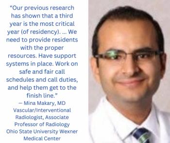
Medical errors are the third most common cause of patient mortality in the United States, following cancer and cardiovascular disease, affecting more than 251,000 patients annually(1).
Errors in diagnostic interpretative imaging are a key contributor with an estimated day-to-day error rate of 3 percent-to-5 percent for all studies reported(2). With 1 billion radiographic examinations performed annually worldwide, this would translate into 30 million-to-50 million interpretative errors per year. Given the significant patient implications, it is imperative that we continually attempt to identify and eliminate diagnostic errors as best as possible.
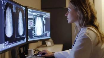
A common diagnostic error is termed “satisfaction of search.” With this error, a radiologist fails to continue to search for additional abnormalities after finding the first one.(3) This error accounts for 22 percent of radiologic errors.
Using checklists is the classic remedy. A helpful checklist is well encompassing, but not too lengthy. It can reduce errors of omission by reminding the radiologist to take a second look at certain imaging features, particularly for “do-not-miss” diagnoses(3). Additionally, working smarter by simply being mindful of diagnoses that oftentimes present in combination, such as multiple fractures, foreign bodies or vascular occlusions, can help reduce the likelihood of finishing a report too early before all of the abnormalities have been identified(4).
Under-reading is another common diagnostic error, constituting up to 42 percent or more of all radiologist errors(3). It occurs when an abnormality is present on the image but is missed and not recorded in the final report. Often, the abnormality is easily identifiable in retrospect, but it was simply not perceived at the time of primary image interpretation(3).
While there is no definitive solution, several strategies can help reduce the frequency of this type of error. For example, image enhancement and post-processing tools can help optimize images and aid in reducing under-reading errors. An example of this is bone subtraction software being used to reduce anatomic noise and help visualize pulmonary nodules on chest radiographs. In addition to enhancing the image and reducing anatomic noise, artificial intelligence is becoming increasingly more useful at identifying abnormalities in the image itself. Computer-aided detection can help reduce the likelihood of missing some radiologic abnormalities, particularly in mammography and lung nodule detection on CT(2).
The third key error that affects imaging care is failure of effective communication, which is often a key driver of patient adverse outcomes. Effective communication between the radiologist and the referring physician relies on clear and concise reports delivered in an appropriate and timely manner.
To improve the overall structure of the report, the American College of Radiology (ACR) Reporting and Data Systems (RADS) provides a standardized framework for reporting on imaging findings. This framework can reduce the variability between reports on a diverse range of disease processes. In addition to the content of the report itself, optimal time frame for communication is also an important aspect to consider as a source of error(5). To this end, the ACR Actionable Reporting Work Group categorized findings in terms of optimal time frame for communication from most-to-least urgent, which provide guidance regarding the response times to improve interdisciplinary communication and patient outcomes.
Identifying major diagnostic errors and potential strategies to reduce their occurrence is a key step towards improving radiologic care. However, perhaps the most important factor to consider is the inclusion of meta-awareness in the education of future radiologists. Providing the next generation of radiologists with an understanding of their own limitations and biases may have the greatest long-term impact on reducing the frequency of diagnostic errors.
We must never lose sight of the fact that, as humans, we are prone to error despite our best intentions, and by acknowledging our fallibility and working diligently to change them, we will continue to improve.
Sources:
1. Makary MA, Daniel M. Medical error-the third leading cause of death in the US. Bmj 2016:i2139. doi:10.1136/bmj.i2139.
2. Brady AP. Error and discrepancy in radiology: inevitable or avoidable? Insights into Imaging. 2016;8:171–82.
3. Bruno MA, Walker EA, Abujudeh HH. Understanding and Confronting Our Mistakes: The Epidemiology of Error in Radiology and Strategies for Error Reduction. RadioGraphics 2015;35:1668–76. doi:10.1148/rg.2015150023.
4. Lee CS, Nagy PG, Weaver SJ, Newman-Toker DE. Cognitive and System Factors Contributing to Diagnostic Errors in Radiology. American Journal of Roentgenology 2013;201:611–7. doi:10.2214/ajr.12.10375.
5. Waite S, Scott JM, Drexler I, Martino J, Legasto A, Gale B, et al. Communication errors in radiology – Pitfalls and how to avoid them. Clinical Imaging 2018;51:266–72. doi:10.1016/j.clinimag.2018.05.025.
Newsletter
Stay at the forefront of radiology with the Diagnostic Imaging newsletter, delivering the latest news, clinical insights, and imaging advancements for today’s radiologists.


