
- AI
- Molecular Imaging
- CT
- X-Ray
- Ultrasound
- MRI
- Facility Management
- Mammography
Could a Deep Learning Model for Mammography Improve Prediction of DCIS and Invasive Breast Cancer?
Artificial intelligence (AI) assessment of mammography images may significantly enhance the prediction of invasive breast cancer and ductal carcinoma in situ (DCIS) in women with breast cancer, according to new research presented at the Society for Breast Imaging (SBI) conference.
Previously validated for breast cancer risk prediction at multiple facilities, a deep learning algorithm for mammography recently demonstrated over 25 percent higher area under the curves (AUCs) for predicting ductal carcinoma in situ (DCIS) and invasive breast cancer in comparison to traditional models.
In the retrospective study, recently presented at the Society for Breast Imaging (SBI) conference, researchers compared a deep learning model (Mirai), based solely on mammography image assessment, to the eighth version of the Tyrer-Cuzick (TC) and the Breast Cancer Risk Assessment Tool (BRCAT) for the prediction of DCIS and invasive breast cancer.
The cohort was comprised of 1,201 women with breast cancer (median age of 60) with 882 cases of invasive breast cancer and 319 cases of DCIS. The researchers noted that 86.7 percent of the cohort had a personal history of breast cancer and 78.9 percent had a family history of breast cancer. Additionally, 79 percent of the study population were post-menopausal women and 59.8 percent did not have dense breasts, according to the study. The racial makeup of the cohort was largely White (81.8 percent) with Asian women comprising 5.5 percent women. Black women accounting for 4.7 percent and people of other races making up 5.9 percent of the group.


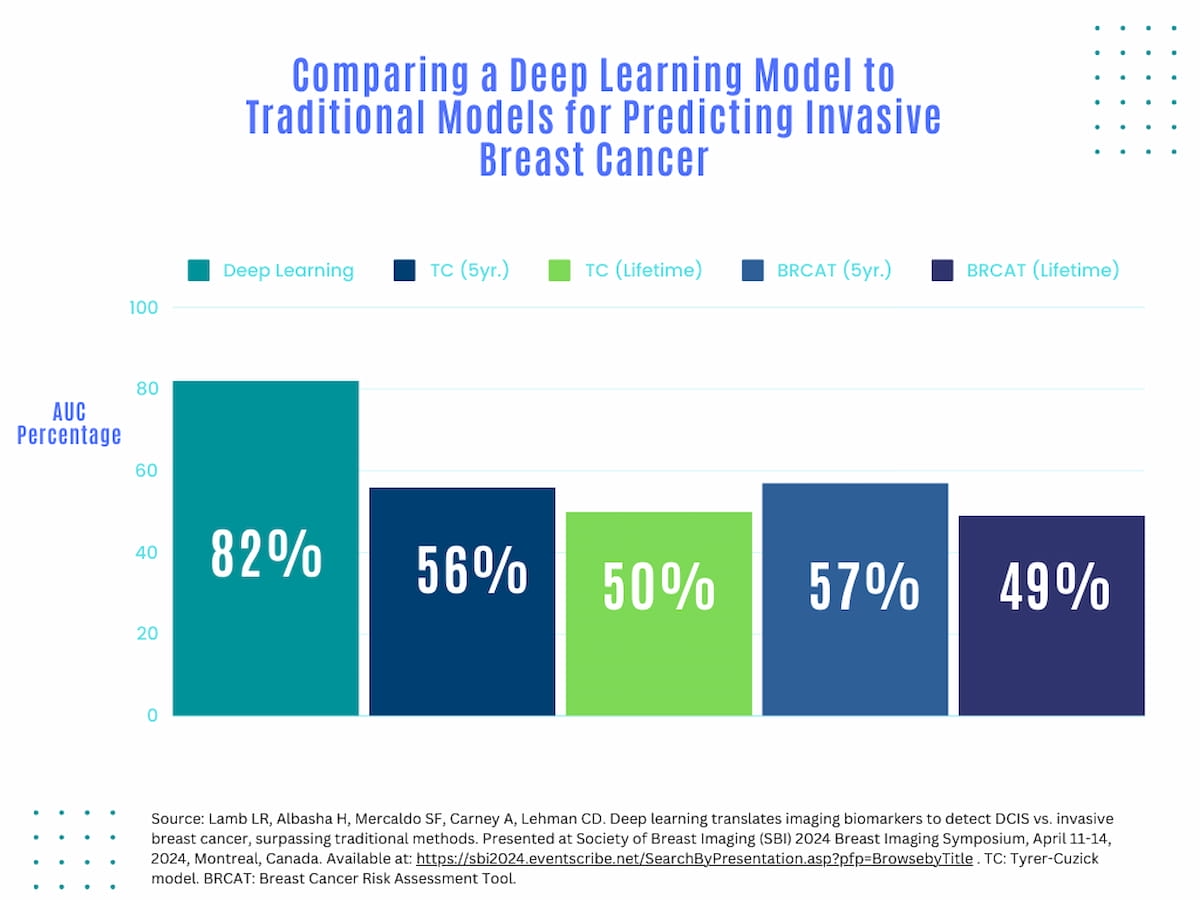
For invasive breast cancer prediction one year after an index mammogram, the deep learning model showed a greater than 25 percent higher AUC (82 percent) in contrast to five-year and lifetime models for the TC system (56 percent and 50 percent respectively) and the BRCAT (57 percent and 49 percent respectively).
Within one year of the index mammogram, the study authors found that the deep learning model had a greater than 25 percent higher AUC (84 percent) for DCIS prediction than five-year and lifetime models for TC system (57 percent and 50 percent respectively) and the BRCAT (55 and 49 percent respectively).
For invasive breast cancer prediction one year after an index mammogram, the researchers saw similar results with the deep learning model showing a greater than 25 percent higher AUC (82 percent) in contrast to five-year and lifetime models for the TC system (56 percent and 50 percent respectively) and the BRCAT (57 percent and 49 percent respectively).
‘Mammograms contain highly predictive biomarkers of breast cancer risk not identified by traditional models,” noted lead study author Leslie R. Lamb, M.D., MSc, who is affiliated with Massachusetts General Hospital (MGH) in Boston.
Noting the challenges of low AUCs with other risk-based screening tools in breast cancer prediction, the study authors suggested the deep learning model has the potential to bolster prediction for DCIS and invasive breast cancer.
“A (deep learning) model using screening mammography alone can improve risk discrimination accuracy in identifying both DCIS and invasive disease compared to traditional modern risk models,” noted Dr. Lamb, who is affiliated with Massachusetts General Hospital in Boston.
In regard to study limitations, the researchers acknowledged that the racial makeup of the cohort (over 80 percent White) may limit extrapolation of the findings to broader populations. They also conceded that one vendor system was used for all mammography exams and that the mammograms used to assess the deep learning model came from MGH where the model was originally developed.
Reference
1. Lamb LR, Albasha H, Mercaldo SF, Carney A, Lehman CD. Deep learning translates imaging biomarkers to detect DCIS vs. invasive breast cancer, surpassing traditional methods. Presented at Society of Breast Imaging (SBI) 2024 Breast Imaging Symposium, April 11-14, 2024, Montreal, Canada. Available at: https://sbi2024.eventscribe.net/SearchByPresentation.asp?pfp=BrowsebyTitle .


 Technology Provide a Viable Alternative to X-Rays for Aortic Procedures?")


CT Study: AI Algorithm Comparable to Radiologists in Differentiating Small Renal Masses
May 14th 2024An emerging deep learning algorithm had a lower AUC and sensitivity than urological radiologists for differentiating between small renal masses on computed tomography (CT) scans but had a 21 percent higher sensitivity rate than non-urological radiologists, according to new research.


Current Insights and Emerging Roles for Contrast-Enhanced Mammography
May 10th 2024In a recent lecture at the 2024 ARRS Annual Meeting, Jordana Phillips, MD, discussed the role of contrast-enhanced mammography in staging breast cancer, evaluating response to neoadjuvant chemotherapy and recalls from screening.


Study Finds High Concordance Between AI and Radiologists for Cervical Spine Fractures on CT
May 6th 2024Researchers found a 98.3 percent concordance between attending radiology reports and AI assessments for possible cervical spine fractures on CT, according to new research presented at the 2024 ARRS Annual Meeting.

FDA Clears AI-Powered Qualitative Perfusion Mapping for Cone-Beam CT
May 6th 2024Reportedly validated in more than 10 clinical trials, the AngioFlow perfusion imaging software enables timely identification of brain regions with cerebral blood flow reduction and those with significant hypoperfusion.