|Articles|December 1, 2008
CTA evaluates coronary stent patency
Author(s)James Brice
Cohort studies presented in a scientific session Sunday showed progress toward employing coronary CT angiography to evaluate the patency of coronary artery stents.
Advertisement
Cohort studies presented in a scientific session Sunday showed progress toward employing coronary CT angiography to evaluate the patency of coronary artery stents.
Building on research presented in the two preceding years, the results covered during this year's sessions indicate that the protocols have improved for examining stent patency with 64-slice or dual-source CT. Promising findings from this still-preliminary work justify their adoption as alternatives to x-ray angiography by academic sites and will lead to more research on larger patient populations to move them into general use.
Results presented by Dr. Mariko Ehara of Toyohashi Heart Center in Japan identified the most likely sites for coronary stent fractures in a study of 272 consecutive patients and 430 stented lesions. Ehara's findings suggest that radiologists and cardiologists look to the right coronary artery, especially the ostium, and sites of multiple stent placement or placement in bended arteries as the most likely locations of stent fracture.
Overall, fractures were identified in 35 (8%) of the stents based on an evaluation using a 64-slice CT scanner. Ehara broke down the incidence of fracture:
- Graft: one of two (50%)
- Right coronary artery: 26 of 150 (17.3%)
- Left anterior descending artery: eight of 173 (4.6%)
- Left coronary artery: zero of 84 (0%)
- Left main: zero of 21 (0%)
Ehara also identified fractures in 32 of 337 stents (9.5%) placed in proximal locations and three of 93 stents (3.2%) located in a distal branch.
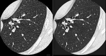
Multislice CT was particularly useful for uncovering fractures because of its ability to depict 3D stent structure and in-stent restenosis simultaneously. It also offered the opportunity to perform repeat evaluations noninvasively.
Radiologist Dr. Lingyan Kong of the Peking Union Medical College Hospital in China presented two studies investigating the value of dual-source CTA for detecting in-stent restenosis. Her prospective study of 30 patients found that the dual-source approach correctly detected 12 of 14 in-stent stenoses. The sensitivity, specificity, positive predictive value, and negative predictive value of the approach were 85.4%, 95.7%, 85.4%, and 95.7%, respectively.
Results from Kong's second study of 78 patients illustrated the importance of stent diameter in the ability to visualize in-stent vessel lumina with dual-source CT. Using a five-point scale ranging from 1 for excellent to 5 for poor image quality, an experienced blinded reader judged the overall ability of dual-source CT to visualize in-stent vessel lumina at 1.6 (±0.6).
The scores closely correlated with stent diameter, with the larger stents producing the better scores. Image quality was relatively poor with the presence of calification.
Advertisement
Related Content


Advertisement
Advertisement
Trending on Diagnostic Imaging
1
The Hidden Social Price of Remote Work in Radiology
2
Addressing Challenges in Radiology Reporting
3
Study: PSMA PET/CT Reduces Biopsy Rate by Nearly 50 Percent for Men with Equivocal or Non-Suspicious Prostate mpMRI
4
FDA Clears AI-Powered Software for Improving Low-Contrast CT Detection
5