CAD expands to aid imaging in niches throughout body
When computer scientists began seriously plumbing the medical mind 30 years ago for the diagnostic process buried within, physicians worried about their future place in the grand scheme of healthcare. Their concern soon waned as the focus of artificial intelligence projects narrowed from all diseases to a few and now, finally, to the task of identifying suspicious lesions on digital images.
When computer scientists began seriously plumbing the medical mind 30 years ago for the diagnostic process buried within, physicians worried about their future place in the grand scheme of healthcare. Their concern soon waned as the focus of artificial intelligence projects narrowed from all diseases to a few and now, finally, to the task of identifying suspicious lesions on digital images.
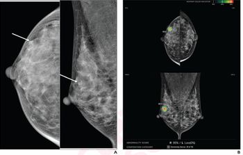
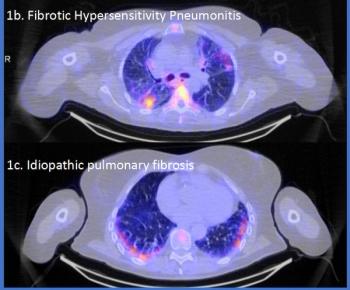
Today computer-aided detection, a moniker that pays homage to the emotionally charged issue of machines participating too much in the diagnostic process, is carving niches in challenging areas of medical interpretation. It took root in mammography, becoming a routine part of digital mammography. It is spreading to breast MR and chest CT with commercial products widely available. Research is driving advances in these and other applications addressing the colon, brain, liver, kidney, bone, and vasculature.
Hologic's subsidiary and CAD pioneer R2 Technology is a major provider of mammography CAD, offering software for use on the Hologic-built Selenia full-field digital mammography system, as well as stand-alone workstations. R2 competitor iCAD supplies CAD mammography products to other vendors, namely Siemens and GE, while providing CAD workstations and software to mammographers.
At the RSNA meeting, both will showcase their latest developments, R2 its most recent ImageChecker and iCAD its SecondLook Digital. The Hologic exhibit will draw special attention to its R2 DigitalNow, which digitizes prior film mammograms, then archives the digital images to PACS for use in a soft-copy viewing environment.
With less than one-fourth of mammography systems converted to digital, growth of full-field digital mammography is expected to continue for years. CAD systems are currently being purchased nearly one-to-one with every digital mammography system purchase, according to iCAD.
iCAD remains well positioned to provide CAD functionality and workflow with all leading PACS and mammography review workstations. In the near term, iCAD plans to unveil a scalable CAD platform to increase sensitivity to masses and microcalcifications with all FFDM systems, while at the same time improving false-positive detection, according to the company. Additionally, iCAD says future releases will increase the amount of clinically relevant information provided about suspicious lesions to help radiologists make diagnoses.
CAD Sciences will feature CAD for breast, prostate, therapy monitoring, and angiography, talking up algorithms released earlier this year for its CAD programs that correct motion artifact in MR breast and prostate imaging, as well as lung CT. Confirma will showcase its CADstream for breast and prostate MRI.
Median Technologies will feature its Lesions Management Solution package of lung, colon, and liver CAD. Medical imaging lab im3D will highlight an algorithm that can shorten the reading time of CT virtual colonography. Riverain Medical will showcase its newly minted OnGuard, a chest x-ray CAD product.
Yet, true to the history of its grand aspirations of the past, CAD still falls short, even in the most restrictive applications. Specificity and sensitivity are below what many prospective customers would like to see in mammography. This was underscored earlier this year by an article published in The New England Journal of Medicine, challenging the diagnostic value of CAD as a second reader of mammograms. Advocates of the technology blasted the report and its conclusions, but none could say CAD mammography was flawless.
Its greatest challenge, a lack of specificity, forces interpreters to recheck too many lesions, slowing the diagnostic process at a time when the need for productivity is becoming more pronounced. Engineers are whittling away at the problem, improving CAD little by little, just as the technology spreads to other applications.
While computer-aided diagnosis may not meet its earliest expectations, computer-aided detection has at least satisfied some very specific clinical needs-and, with advances shown at the RSNA meeting, will contribute even more.
Newsletter
Stay at the forefront of radiology with the Diagnostic Imaging newsletter, delivering the latest news, clinical insights, and imaging advancements for today’s radiologists.

