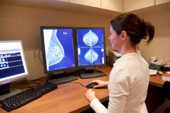
Breast Cancer Risk Increase with Repeat Imaging
CHICAGO - Breast cancer risk estimates increase with frequent CT scans, particularly among younger patients, researchers found.
CHICAGO - Between 1980 and 2006, the per capita exposure to radiation from diagnostic imaging in America increased six-fold. And now, new research suggests that breast cancer risk estimates increase with frequent CT scans, particularly among younger patients, according to a
“We already know there is an increased risk of cancer after an exposure to radiation that's in the same range as is delivered by imaging, particularly CT exams,” said Ginger Merry, MD, breast imaging fellow at Chicago's Prentice Women's Hospital, Northwestern Memorial Hospital and leader of the study.
Since breast tissue is known to be particularly radiosensitive, researchers aimed to examine the impact of increased imaging on radiation exposure and resulting breast cancer risk. Merry and her colleagues performed a retrospective study of about 250,000 women enrolled in a large integrated health care system between 2000 and 2010.
“We gathered data on all examinations that could deliver radiation to the breast tissue, including CT, radiography, fluoroscopy and nuclear medicine, performed by injecting a radiopharmaceutical into the blood stream. Over the 10 year period, we looked at their cumulative radiation exposure,” Merry said.
Using smaller data samples, Merry and her team were able to estimate the average radiation dose given during each examination and calculate the associated breast cancer risk. Then, they compared the imaging-risk to a baseline risk, beginning 10 years after the exposure.
"We found that the estimated breast radiation doses from CT were highly variable across patients, with the highest doses coming from multiple-phase cardiac and chest CT examinations, where successive images of the organ being studied are captured," said senior author Rebecca Smith-Bindman, MD, of the University of California, San Francisco.
More troubling, pediatric doses were more similar than expected to doses administered to adults, and when repeated CT examinations were performed at an early age, young women had an increased risk of breast cancer.
“Younger women are at the highest risk due to increased radiosensitivity of breast tissue and their longer lifespans, and therefore a longer time period in which to develop breast cancer,” Merry said.
For example, if a girl with no other familial or genetic risk factors of breast cancer were to have two chest CT scans at age 15, her risk at age 25 of developing breast cancer in the next 10 years would double. The longer the time since the exposure, the greater the risk of cancer.
To lower imaging-associated breast cancer risk, radiologists should carefully evaluate the need for imaging and monitor radiation doses given with each exam, Smith-Bindman said. Radiation exposure could also be minimized by decreasing the amount of multi-phase protocols whenever possible.
"If imaging is truly indicated, then the risk of developing cancer is small and should not dissuade women from getting the test they need," she said. "On the other hand, a lot of patients are undergoing repeat chest and cardiac CT, many of which aren't necessary. Women, and particularly young women, should understand there is a small but real potential risk of breast cancer associated with cardiac and chest CT, and the risk increases with the number of scans."
Newsletter
Stay at the forefront of radiology with the Diagnostic Imaging newsletter, delivering the latest news, clinical insights, and imaging advancements for today’s radiologists.


