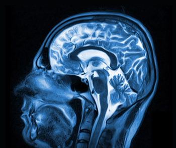
Brain Alterations Seen Via MRI in COVID-19-Positive Technologist
Technologist positive for the virus experienced a loss of smell and altered sense of taste.
MRI images of a young radiology technologist positive for COVID-19 have revealed the virus might find its way into the brain via the olfactory pathway, causing those sensory nerves to malfunction.
Through results published in the May 29
This is only report of this kind, according to the team, led by Letterio Politi, M.D.
“To our knowledge, this is the first report of in vivo human brain involvement in a patient with COVID-19 showing a signal alternation compatible with viral brain invasion in a cortical region (i.e., posterior gyrus rectus) that is associated with olfaction,” they wrote. “Alternative diagnoses (e.g., status epilepticus, posterior reversible encephalopathy syndrome-like alterations, other viral infections, and anti-N-methyl-D-aspartate receptor encephalitis) are unlikely given the clinical context.”
Instead, they said, the results pointed to the possibility that SARS-CoV-2, the virus that causes COVID-19, could use the olfactory pathway to invade the brain. Cerebrospinal fluid and pathology studies must be used to confirm this notion.
The technologist, who had been working in the IRCCS emergency department and had contact with COVID-19-positive patients, presented to the hospital on March 15 with anosmia. After experiencing a mild cough for a day, she developed severe anosmia and a distorted sense of taste. Still, she did not have a fever, nor did she have trauma, seizure, or a hypoglycemic event.
After three days, a nasal fibroscopic evaluation produced unremarkable results, and non-contrast chest and maxillofacial CT results were negative. A brain MRI was performed the same day. On both 3D and 2D fluid-attenuated inversion recovery images (FLAIR), a cortical hyperintensity was apparent in the right gyrus rectus, as well as a subtle hypersensitivity in the olfactory bulbs.
After 28 days, a follow-up MRI revealed that the signal alteration in the cortex had completely disappeared, and the olfactory bulbs were thinner and slightly less hyperintense. The technologist also recovered from anosmia.
Based on these findings and those from other patients, the team said, it maybe possible that imaging changes may not always be present in patients with COVID-19 -- or they might be limited to the very early phase of the disease. But, the role of anosmia could be more significant.
“Anosmia can be the predominant COVID-19 manifestation,” they said. “And, this should be considered for the identification and isolation of patients with infection to avoid disease spread.”
Newsletter
Stay at the forefront of radiology with the Diagnostic Imaging newsletter, delivering the latest news, clinical insights, and imaging advancements for today’s radiologists.


