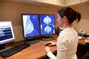
Better QA procedures help eliminate breast errors
Developing a structured and rigorous peer-review quality assurance process that involves ongoing case presentations, open discussion, and consensus opinions can help to decrease perception errors and improve the interpretive skills of breast imagers, according to radiologists at Beth Israel Deaconess Medical Center in Boston.
Developing a structured and rigorous peer-review quality assurance process that involves ongoing case presentations, open discussion, and consensus opinions can help to decrease perception errors and improve the interpretive skills of breast imagers, according to radiologists at Beth Israel Deaconess Medical Center in Boston.
"Between 10% and 30% of breast cancers are missed on mammography, and up to half of all cancers are identified retrospectively on a prior screening mammogram," said Dr. Valerie Fein-Zachary, radiologist and the principal author of an education exhibit presented at RSNA 2009. "To reduce errors of perception and interpretation, we must increase our understanding of why these errors occur."
The identification of a missed breast cancer is subjective and controversial. She defines a missed breast cancer as a subset of false-negative cases where a consensus of reviewers in a well-organized QA environment identifies a suspicious finding on imaging (mammogram, ultrasound, or MRI). She defines interval cancers as those occurring within one year of a negative breast imaging examination.
Beth Israel Deaconess has six ways of collecting possible cases of missed breast cancer:
- the annual medical audit of all screening and diagnostic mammograms, which identifies newly diagnosed cancers
- the online radiology peer-review system, for which each radiologist reviews at least 5% of his/her practice volume and posts the cases online with one of four codes (agreeing or disagreeing with the interpretation)
- the biweekly multidisciplinary breast conference, at which 90% of newly diagnosed cases of breast cancer are discussed
- the weekly breast radiology-pathology conference of interesting and discordant cases
- diagnostic and screening cases where a breast imager identifies a QA issue
- the hospital online QA program
All successful QA programs have a designated QA radiologist, Fein-Zachary said. The QA breast radiologist collects the data and cases for QA presentations, including missed breast cancers. The person is trusted to maintain confidentiality, and every month or two, four to six cases are presented to the breast imagers for review, including at least one "normal" case. The presentation of the cases includes the target exam and a comparison, if available. It may also include subsequent follow-up studies.
The breast imagers, fellows, and senior residents complete evaluation sheets, with exam dates and breast peer-review QA codes for each study. The group has one minute to evaluate each study, and the presenter continues through the sequence, providing appropriate history. A radiologist highlights the main teaching points, and a consensus is reached.
"The educational value of discussing missed breast cancers in a confidential, nonthreatening environment with staff, fellows, and residents cannot be overstated," she said. "Done properly, we have found no problem with the two major traditional challenges to QA programs: the fear of legal repercussions and the difficulty of discussing personal errors in a group setting."
Although cases under discussion are anonymous and remote, some radiologists appreciated prior notification of cases in which they were involved, she said. Lectures with legal and risk management departments also help to alleviate tensions.
Fein-Zachary acknowledges that the major impediment to instituting this type of breast peer-review QA process is time. To create a meaningful teaching experience, the QA radiologists must review cases and prepare them in advance. Consensus peer-review requires at least three radiologists, which may not be feasible in smaller practices. A more practical method of identifying missed cancers may be to cross-reference the database of breast imaging patients with state-wide cancer registries, she said. This would be particularly helpful for practices without electronic medical records or where surgical follow-up is not at the same institution.
Newsletter
Stay at the forefront of radiology with the Diagnostic Imaging newsletter, delivering the latest news, clinical insights, and imaging advancements for today’s radiologists.


