AI Can ID Lung Cancer on X-rays in Healthy Patients As Well As Radiologists
A commercially available deep learning algorithm performed comparably to the individual radiologist when assessing patients at low risk for the disease.
Using artificial intelligence (AI) to detect lung cancer on X-rays in the healthy general population can be just as effective as relying on a radiologist’s interpretation, potentially freeing providers up to focus on other studies, according to a new study.
Investigators from Seoul National University College of Medicine in Korea tested a deep learning algorithm that is already commercially available from Lunit with patients who participated in a screening program at Seoul National University Hospital Health System Gangnam Center between January 2008 and December 2012. Performance, they found, was equivalent to a radiologist alone.
“A deep learning algorithm detected lung cancer nodules on chest radiographs with a performance comparable to that of radiologists, which will be helpful for radiologists in healthy populations with a low prevalence of lung cancer,” said the team led by Chang Min Park, M.D., Ph.D., associate professor of radiology at Seoul University Hospital.
The team published their findings in the Sept. 22
Related Content:
Recently, AI has been gaining a foothold in lung cancer detection with X-ray in at-risk patient populations, Park’s team said. But, they set out to determine whether those same tools can be used effectively in healthy groups.
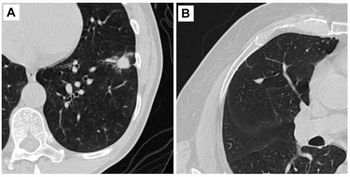
To make that assessment, they used a validation set comprised of 10,285 chest X-rays with 10 visible lung cancers. With this set, the algorithm had an area under the curve of 0.99 and comparable sensitivity to the individual radiologist – 90 percent and 60 percent, respectively. Their screening cohort included 100,525 chest X-rays with 47 visible lung cancers. The algorithm performed similarly, they said, with an area under the curve of 0.97 and sensitivity of 83 percent. For both sets, the false positive rate was roughly 3 percent.
Not only did these findings show equivalency between AI and radiologist performance, the team said, but the indicated the algorithm could be applied to wide patient populations.
“We performed our study in a real-world setting, using a real-world health screening population,” they said. “Therefore, we believe that the algorithm analyzed in our study could be reasonably applied for the detection of lung cancers on chest radiographs in a health screening population with an average risk of lung cancer.”
Implementing the algorithm, the team said, could reduce the number of simple mistakes or perceptual errors that can sometimes pop up with radiologists who have less experience.
In an accompanying editorial, Samuel G. Armato, III, Ph.D., associate professor of radiology at the University of Chicago, did note that Park’s team used a pre-determined output threshold for their study, but any subsequent work most likely require system re-training. In addition, he said, the team did not include the quantified results extracted from lesion localization maps returned by the algorithm.
"Knowledge gained from such an assessment would benefit the medical imaging research community,” he said.
Ultimately, the team acknowledged that, even with these findings, additional work is necessary.
“To generalize the clinical use of the deep learning algorithm in a screening program of the general population,” they said, “further studies covering a variety of races and medical environments will be needed.”
For more coverage based on industry expert insights and research, subscribe to the Diagnostic Imaging e-newsletter here .
Newsletter
Stay at the forefront of radiology with the Diagnostic Imaging newsletter, delivering the latest news, clinical insights, and imaging advancements for today’s radiologists.


