4D-Fueled AI with DCE-MRI Improves Breast Lesion Characterization
New method can help providers distinguish between benign and malignant breast lesions.
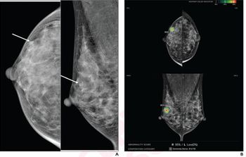
Radiologists can classify breast lesions more accurately if they use artificial intelligence algorithms fueled by 4D data captured with dynamic contrasted-enhanced MRI (DCE-MRI), new research has found.
In a study published on Feb. 24 in
“Incorporating 4D information in DCE-MRI by feature MIP in deep transfer learning demonstrated superior classification performance compared with using MIP images as input in the task of distinguishing between benign and malignant breast lesions,” said the team led by doctoral student Qiyuan Hu, noting that their method outperforms the existing MIP strategy that only uses one post-contrast subtraction image.
This new method is the second attempt by University of Chicago researchers to use 4D information from DCE-MRI. The first iteration, dubbed image MIP, collapsed suspicious 4D DCE-MRI areas of interest from second post-contrast subtraction images down into 2D MIP, and, its results were better than those rendered by 3D-based methods.
But, Hu’s team took it a step further.
“Instead of collapsing the volumetric information at the image level to form MIP images, we do so at the feature level by taking the maximum of CNN features along the axial dimension for a given lesion directly within the deep neural network architecture,” they said.
To gather more substantive temporal information, this method – called feature MIP – incorporates subtraction images from four dynamic points in the DCE sequence that are, then, put into red, green, and blue CNN channels. The team postulated that this new method might perform better in distinguishing between benign and malignant breast lesions because it might better leverage DCE-MRI’s volumetric information.
They set out to prove their hypothesis by using a retrospective dataset of 1,990 distinct lesions – 1,494 malignant and 496 benign – from 1,979 women. With a training and validation set of 1,455 lesions gathered between 2015 and 2016, Hu’s team trained linear support vector machine classifiers to characterize lesions as benign or malignant based on features extracted from the CNN.
Based on their analysis, the team determined that, on an independent 535-lesion test set, image MIP and feature MIP produce areas under the curve of 0.91 and 0.93, respectively, with the feature MIP method outperforming image MIP.
The team said they plan to expand this method to T2-weighted and diffusion-weighted MRI sequences in multi-parametric MRI, using images gathered from other medical centers and groups. Doing so, they concluded, will allow them to evaluate how their method performs across manufacturers, protocols, and patient groups.
For more coverage based on industry expert insights and research, subscribe to the Diagnostic Imaging e-Newsletter
Newsletter
Stay at the forefront of radiology with the Diagnostic Imaging newsletter, delivering the latest news, clinical insights, and imaging advancements for today’s radiologists.


